10 Key Points from the 2013 ESC Guidelines on the Management of Stable Coronary Artery Disease

10 Key Points from the 2013 ESC   Guidelines on the Management of Stable Coronary Artery Disease


These guidelines should be applied to patients with stable known or suspected coronary artery disease (SCAD). This condition encompasses several groups of patients: (i) those having stable angina pectoris or other symptoms felt to be related to coronary artery disease (CAD) such as dyspnoea; (ii) those previously symptomatic with known obstructive or non-obstructive CAD, who have become asymptomatic with treatment and need regular follow-up; (iii) those who report symptoms for the first time and are judged to already be in a chronic stable condition (for instance because history-taking reveals that similar symptoms were already present for several months). Hence, SCAD defines the different evolutionary phases of CAD, excluding the situations in, which coronary artery thrombosis dominates clinical presentation (acute coronary syndromes).

The following are 10 key points from the 2013 European Society of Cardiology guidelines for the management of stable coronary artery disease (CAD), with emphasis since the last guidelines publication in 2006:
1. There are different underlying mechanisms of stable known or suspected CAD (SCAD). These include fixed or dynamic plaque-related obstruction of epicardial arteries, focal or diffuse spasm of normal or plaque-disease arteries, microvascular dysfunction, and left ventricular dysfunction caused by prior acute myocardial necrosis and/or hibernation. These mechanisms allow for consideration of ‘microvascular dysfunction and coronary vasospasm in diagnostic and prognostic algorithms.’
2. Coronary computed tomography angiography (CTA) may be considered an alternative to ischemia testing, especially in patients with chest pain symptoms with low to intermediate pretest probability. While CTA should not be overused, its very high negative predictive value can provide reassurance in select circumstances.
3. Although promising and valuable in offering information on both overall cardiac anatomy and function in the same examination, magnetic resonance coronary angiography is still regarded as a research tool and is not officially recommended in the diagnostic evaluation of SCAD.
4. Cardiac rehabilitation, commonly offered after myocardial infarction or recent coronary intervention, should be considered in all patients with SCAD.
5. The treatment of microvascular angina includes optimal coronary risk factor control and traditional anti-ischemic therapy. Beta-blockers are recommended as first-line therapy, given their role in relieving effort-related angina.
6. Patients with a high pretest probability for CAD and/or severe symptoms may benefit from early invasive coronary angiography without noninvasive risk stratification.
7. Fractional flow reserve (FFR), intravascular ultrasound, and optical coherence tomography are methods of intracoronary assessment of coronary artery stenosis severity. The measurement of FFR during adenosine infusion may identify functionally significant stenosis and should be used frequently. A patient with a stenosis and an FFR >0.80 should not be revascularized.
8. While it has been well established that there is genetic variation to the response to antiplatelet therapy (especially clopidogrel) in patients with acute coronary syndrome or myocardial infarction, there are no established recommendations to perform genetic testing to guide treatment with antiplatelet therapy in patients with SCAD.
9. Ranolazone, a selective inhibitor of late sodium current, has anti-ischemic and metabolic properties. It is useful as add-on treatment for the management of stable angina in patients inadequately controlled by first-line therapy, and does not impact heart rate or blood pressure.
10. In light of the results of the FREEDOM trial, coronary artery bypass grafting may be the preferred revascularization strategy in diabetic patients with multivessel disease.


Source:   Cardiosource


Are ICDs Useful in Preventing SCD in Children and Adolescents with HCM?

Hypertrophic cardiomyopathy (HCM) is the most common cause of sudden cardiac death (SCD) in the young. The availability of  implantable cardioverter-defibrillators (ICDs) over the past decade for HCM has demonstrated the potential for sudden death prevention, predominantly in adult patients. In children and adolescents with hypertrophic cardiomyopathy , ICDs terminated life-threatening ventricular tachyarrhythmias, but frequently led to device-related complications.                                                                                                                                                                                                                                                                                                                                                                                                                         The multicenter, international registry study, published on April 1 in the Journal of the American College of Cardiology, looked at 224 patients with HCM under age 20, who were at high risk for sudden cardiac death (SCD), and who received ICDs for primary or secondary prevention. Results showed ICDs were activated appropriately to terminate ventricular tachycardia or ventricular fibrillation in 43 of 244 patients (19 percent) over a mean of 4.3 ± 3.3 years. Intervention rates were 4.5 percent per year overall, 14.0 percent per year for secondary prevention after cardiac arrest, and 3.1 percent per year for primary prevention on the basis of risk factors.

In addition, the primary prevention discharge rate terminating ventricular tachycardia or ventricular fibrillation was the same in patients who underwent implantation for 1, 2, or ≥3 risk factors (12 of 88 [14 percent], 10 of 71 [14 percent], and 4 of 29 [14 percent], respectively, p = 1.00). Extreme left ventricular hypertrophy was most frequently associated with appropriate interventions.  However, ICD-related complications, particularly inappropriate shocks and lead malfunction, occurred in 91 patients (41 percent) at 17 ± 5 years of age.

The authors support the current risk stratification strategy for identifying patients with HCM susceptible to life-threatening ventricular tachyarrhythmias, and underscore an important role for SCD prevention (with ICDs). However, since the rate of device complications adds a measure of complexity to ICD decisions in this age group, the authors conclude that it is important to balance considerations for the preservation of life using ICDs against the possibility of device-related complications that may be anticipated with implantation so early in life.

Source:

Maron BJ, et al. Prevention of Sudden Cardiac Death with Implantable Cardioverter-Defibrillators in Children and Adolescents with Hypertrophic Cardiomyopathy. J Am Coll Cardiol. 2013;61(14):1527-1535.


Noncompaction Cardiomyopathy (NCCM)

 Isolated non-compaction cardiomyopathy (INCCM) and its typical echocardiographic appearance were first described in 1984 by Engberding and Bender.  INCCM is a heart-muscle disorder that is still little known among physicians.  In a study of children with primary cardiomyopathy of all types, NCCM was present in 9.2%. It was thus the third most common type of primary cardiomyopathy, after dilated cardiomyopathy (DCM) and hypertrophic cardiomyopathy.

Noncompaction of the ventricular myocardium is a cardiomyopathy thought to be caused by arrest of normal embryogenesis of the endocardium and myocardium. This abnormality is often associated with other congenital cardiac defects, but it is also seen in the absence of other cardiac anomalies.  Clinical manifestations are highly variable, ranging from no symptoms to disabling congestive heart failure, arrhythmias, and systemic thromboembolism. Echocardiography has been the diagnostic procedure of choice, but the correct diagnosis is often missed or delayed because of lack of knowledge about this uncommon disease and its similarity to other diseases of the myocardium and endocardium.

 Embryology and Development:

During early embryonic development, the myocardium is a loose network of interwoven fibers separated by deep recesses that link the myocardium with the left ventricular cavity. Gradual “compaction” of this spongy meshwork of fibers and intertrabecular recesses, or “sinusoids,” occurs between weeks 5 and 8 of embryonic life, proceeding from the epicardial to endocardium and from the base of the heart to the apex. The coronary circulation develops concurrently during this process, and the intertrabecular recesses are reduced to capillaries.

The normal process of trabeculation appears to involve secretion of neuregulin growth factors from the endocardium and may also involve angiogenesis factors, such as vascular endothelial growth factor and angiopoietin.

Noncompaction of the ventricular myocardium (NVM) is an uncommon finding. It is thought to be caused by arrest of the normal process of endomyocardial morphogenesis.

NCCM was first described in association with other congenital anomalies, such as obstruction of the right or left ventricular outflow tracts, complex cyanotic congenital heart disease, and coronary artery anomalies. The abnormal compaction process in these cases is not fully understood, but pressure overload or myocardial ischemia preventing regression of the embryonic myocardial sinusoids has been suggested. This process results in the persistence of deep intertrabecular recesses in communication with both the ventricular cavity and the coronary circulation.

Isolated non-compaction cardiomyopathy (INCCM) and its typical echocardiographic appearance were first described in 1984 by Engberding and Bender is characterized by persistent embryonic myocardial morphology found in the absence of other cardiac anomalies to explain the abnormal development. In such cases, the resultant deep recesses communicate only with the ventricular cavity, not the coronary circulation.

The left ventricle is uniformly affected, but biventricular noncompaction has been reported, with right ventricular noncompaction described in less than one-half of patients. Because of difficulty in distinguishing normal variants in the highly trabeculated right ventricle from the pathological noncompacted ventricle, several authors dispute the existence of right ventricular noncompaction.

Both familial and sporadic forms of noncompaction have been described.

In the original report of INCCM, which predominantly involved children, familial recurrence was seen in half of patients.  Familial recurrence was seen in 18% in the largest reported adult population with INCCM, although the authors reported that incomplete screening of siblings may account for the lower percentage compared with the earlier report.

 Epidemiology:

The prevalence of this disease in adults remains unclear. In observational studies, NCCM has been found in 0.014% to 0.26% of all adults referred to an echocardiography laboratory. The incidence of NCCM in the general population has been estimated at 0.05% to 0.25% per year. The diagnosis is presumably often missed, because the disease is still not as well-known as it should be among physicians at large.

Clinical Features and Pathophysiology:

Three major clinical manifestations of noncompaction have been described, including: Heart failure, arrhythmias, and embolic events.

Findings vary among patients, ranging from asymptomatic left ventricular dysfunction to severe, disabling congestive heart failure.

Over two thirds of the patients in the largest series with NCCM had symptomatic heart failure.

Coronary angiography reveals no abnormalities in patients with NCCM, but positron emission tomography (PET) shows a diminished reserve of coronary blood flow in the compact and non-compact myocardial segments of the left ventricle, presumably because of impaired microcirculation. Similar findings are obtained with single photon emission computerized tomography (SPECT).

Depressed ventricular systolic function has been noted in 63% of patients.  Both systolic and diastolic ventricular dysfunction has been described.

Restrictive hemodynamics by cardiac catheterization, as well as an initial presentation of NCCM as a restrictive cardiomyopathy, have been described in children with NCCM.

Left ventricular dysfunction developed in the vast majority, regardless of the presence or absence of symptoms at initial diagnosis.

Furthermore, marked trabeculation can impair the diastolic function of the left ventricle as well, with abnormal relaxation and restricted filling.

The origin of systolic dysfunction in noncompaction is unclear, but it has been suggested that subendocardial hypoperfusion and microcirculatory dysfunction playing roles in ventricular dysfunction and arrhythmogenesis.

These systolic and diastolic disturbances of left ventricular function, if severe enough, can lead to the clinical manifestations of heart failure that are seen in many patients with NCCM.

Arrhythmias are common in patients with ventricular noncompaction. Atrial fibrillation has been reported in over 25% of adults with NCCM. Ventricular tachyarrhythmia has been reported in as many as 47%.   Sudden cardiac death accounted for half of the deaths in the larger series of patients with NCCM.  Paroxysmal supraventricular tachycardia and complete heart block have also been reported in patients with NCCM.

Abnormalities of the resting ECG are found in the majority of patients with NCCM but findings are nonspecific and include left ventricular hypertrophy, repolarization changes, inverted T waves, ST segment changes, axis shifts, intraventricular conduction abnormalities, and AV block. Left bundle branch block has been described in 44% of adult patients with NCCM, but the reported incidence in children was much lower.

Electrocardiographic findings of the Wolff-Parkinson-White syndrome have been described in up to 15% of pediatric patients.

The occurrence of thromboembolic events, including cerebrovascular accidents, transient ischemic attacks, pulmonary embolism, and mesenteric infarction, ranged from 21% to 38%.

Embolic complications may be related to development of thrombi in the extensively trabeculated ventricle, depressed systolic function, or the development of atrial fibrillation.  Of interest, no systemic embolic events were reported in the largest pediatric series with NCCM.

An association between NCCM and facial dysmorphisms, including a prominent forehead, low-set ears, strabismus, high-arching palate, and micrognathia, has been described.

Diagnosis:

Echocardiography is the diagnostic test of choice for NCCM.  Multiple prominent ventricular trabeculations with deep intertrabecular recesses are seen. Color Doppler imaging demonstrates blood flow through these deep recesses in continuity with the ventricular cavity.

The left ventricular apical and inferior wall segments were involved in all patients in an adult population with NCCM studied by echocardiography. The right ventricular apex was involved in 41%.

Depressed left ventricular systolic function was the rule, with a mean calculated ejection fraction of 33% in 28 patients examined. Impairment of diastolic function as assessed by mitral inflow and pulmonary venous flow Doppler was observed in all, including a restrictive filling pattern in 36%.

Prominent trabeculations (although virtually always < 3 in number in normal variants), apical hypertrophic cardiomyopathy, dilated cardiomyopathy, arrhythmogenic right ventricular dysplasia, endocardial fibroelastosis, cardiac metastases, and left ventricular thrombus are important differential diagnostic considerations. Transesophageal echocardiography may be used when transthoracic studies cannot reliably exclude other processes. One report described the use of contrast echocardiography with sonicated albumin in a patient with NCCM. Contrast echocardiography may be helpful when standard echocardiographic image quality is limited or the diagnosis is questionable.

Cardiac magnetic resonance imaging (MRI) is a further method that can be used to diagnose NCCM accurately. Cardiac magnetic resonance imaging has become the method of choice to confirm or rule out left ventricular noncompaction, because echocardiography cannot allow proper visualisation of the apex in some cases.

MRI provides good correlation with echo for localization and extent of noncompaction and is useful in cases with poor echocardiographic image quality.

In addition, the demonstration of differences in MRI signal intensity in noncompacted myocardium may help identify substrate for potentially lethal arrhythmias.

Management:

Treatment for noncompaction of the ventricular myocardium focuses on the 3 major clinical manifestations:  Heart failure, arrhythmias, and systemic embolic events. Standard medical therapy for systolic and diastolic ventricular dysfunction is warranted. Cardiac transplantation has been used for those with refractory congestive heart failure.

The beneficial effects of the beta blocker carvedilol on left ventricular function, mass, and neurohormonal dysfunction in an infant with NCCM have been described.  Because of the frequency of ventricular tachycardia and significant risk of sudden cardiac death and systemic embolism, assessment for atrial and ventricular arrhythmias by ambulatory ECG monitoring should be performed annually. As more information is gathered about NCCM and risk of sudden cardiac death, implantable defibrillator technology may have an expanded role.                                                                                                                                                                                                                                                                                                            Biventricular pacemakers may have a role in the treatment of NCCM patients with heart failure, reduced left ventricular function, and prolonged intraventricular conduction.

Prevention of embolic complications is also an important management issue, and several authors have recommended long-term prophylactic anticoagulation for all patients with ventricular noncompaction whether or not thrombus has been found. Because of the familial association described with noncompaction, screening echocardiography of first degree relatives is recommended. Given the high percentage of associated neuromuscular disorders reported in patients with NCCM, neurological and musculoskeletal evaluations are also recommended.

Prognosis:

Although the prognosis for patients with NCCM varies, nearly 60% of patients described in one large series had either died or undergone cardiac transplantation within 6 years of diagnosis.

Two of 8 in the initially asymptomatic group of this series died during the follow-up period, both having documented sustained ventricular tachycardia and one with sudden cardiac death. Similarly, in a series of 34 adults with NCCM, 47% either died or underwent cardiac transplantation during the follow-up period of 44 months.

The occurrences of systemic emboli, ventricular arrhythmias, and death were considerably lower in the largest pediatric series with NCCM when compared with adults and the initial case series by Chin et al, although nearly 90% of patients followed for up to 10 years developed left ventricular dysfunction.

Oechslin et al reported that certain clinical characteristics were observed significantly more frequently in nonsurvivors compared with survivors with NCCM, including:

1)   –  Higher left ventricular end diastolic diameter on presentation.

2)   –  New York Heart Association class III-IV.

3)   –  Permanent or persistent atrial fibrillation.

4)   –  Bundle branch block.

Patients with these high risk features are candidates for early, aggressive interventions, including consideration of cardioverter-defibrillator implantation and evaluation for transplantation.

Conclusion:

Non-compacted cardiomyopathy is a congenital malformation that can occur as an isolated entity or associated with other pathologies of the heart and can often involve both ventricles. A ratio of noncompacted: compacted wall greater than 3 and involvement of three or more segments are signs of poor prognosis associated with greater clinical deterioration (functional class III/IV) and ventricular arrhythmias.

A high index of suspicion for non-compaction is necessary as the condition may lead to serious heart failure, thromboembolic events, ventricular tachyarrhythmia or death. Early recognition of non-compaction may give better follow-up and management of patients with this condition.

The clinical picture does not provide sufficiently specific evidence to establish the diagnosis. Echocardiography is the diagnostic cornerstone. Treatment should be directed towards prevention and management of heart failure, ventricular arrhythmias and prevention of thromboembolic events. The long-term prognosis of non-compaction cardiomyopathy is poor.

Unresolved issues:

  • Definitive      echocardiographic and CMR criteria for diagnosis.
  • Prevalence of RV      involvement and diagnostic criteria.
  • Whether prognosis      can be improved by early diagnosis-treatment.
  • Comprehension of      the poor genotype-phenotype correlation.

Suggested Readings:

1)   – Weiford B.C., et al; Noncompaction of the Ventricular Myocardium. Circulation 2004, 109:2965-2971.

2)   – Engberding R, et al. Isolated Non-compaction Isolated Cardiomyopathy. Dtsch Arztebl Int. 2010 March; 107(12): 206–213.

3)   – Patil VC, Patil HV. Isolated Non-compaction Cardiomyopathy presented with Ventricular Tachycardia. Hear Views, 2011 Apr-Jun; 12(2):74-78


European Society of Cardiology (ESC) Guidelines on the management of valvular heart disease (version 2012)


 A great number of guidelines have been issued in recent years by the European Society of Cardiology (ESC) as well as by other societies and organizations. Because of their impact on clinical practice, quality criteria for the development of guidelines have been established, in order to make all decisions transparent to the user.

The first step of evaluation of patients with valvular heart disease begins by obtaining a good clinical history. The aim of obtaining a case history is to assess symptoms and to evaluate for associated comorbidity. Essential questions in the evaluation of a patient for valvular intervention should include the following:

• Is valvular heart disease severe?

• Does the patient have symptoms?

• Are symptoms related to valvular disease?

• What are patient life expectancy and expected quality of life?

• Do the expected benefits of intervention (vs. spontaneous outcome) outweigh its risks?

• What are the patient’s wishes?

• Are local resources optimal for planned intervention?

The following are 10 points to remember about these new ESC guidelines on the management of valvular heart disease (VHD):

1. Echocardiography is the key technique used to confirm the diagnosis of VHD, as well as to assess its severity and prognosis. It is indicated in any patient with a murmur, unless no suspicion of valve disease is raised after the clinical evaluation. Transesophageal echocardiography should be considered when transthoracic echocardiography is of suboptimal quality or when thrombosis, prosthetic dysfunction, or endocarditis is suspected.

2. Exercise Stress Testing: The primary purpose of exercise testing is to unmask the objective occurrence of symptoms in patients who claim to be asymptomatic or have doubtful symptoms. Exercise testing has an additional value for risk stratification in AS. Exercise testing will also determine the level of authorized physical activity, including participation in sports.        The use of stress tests to detect coronary artery disease (CAD) associated with severe VHD is discouraged because of their low diagnostic value and potential risks.

Exercise echocardiography may provide additional information in order to better identify the cardiac origin of dyspnea—which is a rather unspecific symptom—by showing, for example, an increase in the degree of mitral regurgitation/aortic gradient and in systolic pulmonary pressures.

3. Coronary angiography is recommended before valve surgery in patients with severe VHD and any of the following: history of CAD; suspected myocardial ischemia; left ventricular (LV) systolic dysfunction; in men aged over 40 years and postmenopausal women; and ≥1 cardiovascular risk factor.

Coronary angiography can be omitted in young patients with no atherosclerotic risk factors (men <40 years and premenopausal women) and in rare circumstances when its risk outweighs benefit, e.g. in acute aortic dissection, a large aortic vegetation in front of the coronary ostia, or occlusive prosthetic thrombosis leading to an unstable hemodynamic condition.

4. In severe aortic regurgitation, surgery is indicated in symptomatic patients, in asymptomatic patients with resting LV ejection fraction (LVEF) ≤50%, and in patients undergoing coronary artery bypass grafting (CABG) or surgery of ascending aorta, or on another valve.

5. Aortic valve replacement is indicated in patients with severe aortic stenosis (AS) and any symptoms related to AS, in patients with severe AS undergoing CABG, surgery of the ascending aorta or another valve, in asymptomatic patients with severe AS and systolic LV dysfunction (LVEF <50%) not due to another cause, and in patients with severe AS and abnormal exercise test showing symptoms on exercise clearly related to AS.

6. Transcatheter aortic valve implantation (TAVI) should only be undertaken with a multidisciplinary ‘heart team’ including cardiologists and cardiac surgeons and other specialists if necessary, and should only be performed in hospitals with cardiac surgery on-site.

7. TAVI is indicated in patients with severe symptomatic AS who are not suitable for AVR as assessed by a ‘heart team,’ and who are likely to gain improvement in their quality of life, and to have a life expectancy of more than 1 year after consideration of their comorbidities.

8. In severe primary mitral regurgitation, mitral valve repair should be the preferred technique when it is expected to be durable. Surgery is indicated in symptomatic mitral regurgitation patients with LVEF >30% and LV end-systolic dimension (LVESD) <55 mm and in asymptomatic patients with LV dysfunction (LVESD ≥45 mm and/or LVEF ≤60%).

9. Percutaneous mitral commissurotomy is indicated in symptomatic mitral stenosis patients with favorable characteristics, and in symptomatic patients with contraindication or high risk for surgery.

10. For valve replacement, a mechanical prosthesis is recommended according to the desire of the informed patient and if there are no contraindications for long-term anticoagulation, and a bioprosthesis is recommended when good quality anticoagulation is unlikely (compliance problems; not readily available) or contraindicated because of high bleeding risk (e.g., prior major bleed, comorbidities, unwillingness, compliance problems, lifestyle, occupation).

Source: 1)- Vahnnian A, et al. Eur heart J 2012; 33:2451-2496; 2)- Mukherjee D.: Cardiosource.


Valve Prosthesis–Patient Mismatch

The concept/phenomenon of valve prosthesis/patient mismatch (VP–PM) was first described in 1978.  All prosthetic heart valves have some degree of VP–PM.

The original paper (in 1978) that described valve prosthesis–patient mismatch (VP–PM) stated that “Mismatch can be considered to be present when the effective prosthetic heart valve area, after insertion into the patient, is less than that of a normal human valve”. It must be noted that all prosthetic heart valves (PHVs) are smaller than normal and thus are inherently stenotic.  The PHV mismatch was stated to be “usually mild to moderate in severity and often of no immediate clinical significance.” However, severe mismatch may lead to significant symptomatic and hemodynamic deterioration and increased mortality.

With the publication of VP–PM, surgeons were more careful and inserted the largest PHV that could be safely inserted. PHVs with improved hemodynamic profile have also been developed. As a result severe VP–PM has become a much less common clinical problem. The overwhelming majority of a large number of scientific publications have related to the aortic valve and not to mitral valve VP–PM.

How should VP–PM be measured?

Different parameters have been evaluated to assess for the severity of VP–PM. The most commonly used measures of valve size is the EOAi.

The EOA is a physiological parameter analogous to the native AVA. The EOA can be measured both invasively and noninvasively using echocardiography/Doppler or magnetic resonance imaging. The most readily and widely available method is echocardiography/Doppler. The accuracy of EOA echocardiographic measurement in the bioprosthetic valve is limited by the same pitfalls that are present in the measurement of the AVA; in particular, the LV outflow tract diameter may be more difficult to measure because of reverberation artifact caused by the prosthetic heart valve, but in these instances, the sewing ring diameter may be a sufficient surrogate. In the bileaflet mechanical valve, the central orifice may produce a high-velocity jet, causing an underestimation of the EOA. Pressure recovery occurs with both bioprosthetic and mechanical heart valves, although the implications of pressure recovery in PHVs have not yet been clarified. The EOAi has a complex relationship with the mean gradient across the aortic valve and PHV.

Assessment of severity of VP–PM

With aortic VP–PM the obstruction to the LV outflow tract is similar to that seen with native AS. Thus, the severity of aortic VP–PM should be assessed by the same criteria as for severe AS. Severe aortic stenosis is defined as mean aortic valve gradient, measured after energy recovery (also called pressure recovery), of ≥50 mm Hg and an AVA index of ≤ 0.6 cm²/m². ); it would be reasonable that this criterion should also be applied to severe VP–PM.


Grading

AVA and EOA, cm2

AVA Index and EOAi, cm2/m2

Mild

>1.5

>0.9

Moderate

>1.0–1.5

>0.6–0.9

Severe

≤1.0

≤0.6

Very severe/critical

≤0.7

≤0.4


When should the severity of VP–PM be determined?

4 phases of physiological healing of mechanical and bioprosthetic PHVs have been described; they are “platelet and fibrin deposition, inflammation, granulation tissue, and finally encapsulation. Long-term device fibrous encapsulation with extension to adjacent tissues adds to structural stability.” Bioprosthetic valves undergo morphological changes of both the tissue material as well as the supporting structures, which may contribute to VP–PM. Valve leaflets become covered by fibrin, platelets, and other cellular material. The matrix of the leaflets undergoes microcalcification as well insulation with plasma materials, causing changes in the matrix structure. These changes may change the resistive properties of leaflet materials. In both mechanical and bioprosthetic valves, a fibrous sheath may also encapsulate the supporting structure of the valve, encroaching on the PHV orifice and also possibly causing valve leaflet or disk immobilization.

For PHVs of the same size, there was a wide range of EOAs. Two echocardiographic/Doppler studies from the Mayo Clinic of patients studied within 1 week of mitral porcine mitral bioprosthesis and of tricuspid mechanical prostheses showed a wide range of gradients and EOAs with the same size of PHV, even in the same brand of PHV.

There are at least several explanations for these findings. 1) PHVs of the same labeled size are not necessarily of precisely the same size. Bioprostheses and other biological valves may also have differences in tissue materials. 2) There are variations from patient to patient with regard to healing changes, hemodynamic conditions, and pressure recovery.

It is best to measure VP–PM early (at 1 week after PHV implantation or at time of hospital discharge) to determine the variations in actual size of the PHV that was implanted in an individual patient. Importantly, VP–PM should also be assessed at 6 to 12 months when the physiological and other morphological changes in the PHV are mostly complete. The severity of VP–PM determined at this time can be expected to determine the long-term impact of VP–PM on patients’ outcomes.

Conclusions:

It must be noted that all prosthetic heart valves (PHVs) are smaller than normal and thus are inherently stenotic.

1) – EOAi should be measured at 1 to 4 weeks or at hospital discharge to evaluate the actual valve size that was implanted. This should also be done at 6 to 12 months to evaluate the severity of VP–PM that will affect long-term outcomes.

2) – The grading of severity of VP–PM should be similar to another common LV outflow tract obstruction, namely, valvular AS. VP–PM can be mild (EOAi >0.9 cm2/m2), moderate (EOAi >0.6 to 0.9 cm2/m2), or severe (EOAi ≤0.6 cm2/m2).

3) – Mild VP–PM, like mild AS, is unlikely to have clinically significant untoward effects. Outcomes with moderate and severe VP–PM should be evaluated separately. Moderate VP–PM is unlikely to reduce survival unless there is progression of valve obstruction, for example, with pannus formation. Severe VP–PM has negative effects on outcomes, but its effect on mortality is still unproven.

4) – Prediction of severity of VP–PM is problematic. The primary goal should be not to prevent VP–PM but rather to prevent severe VP–PM.

5) – Use of the EOAi as a continuous variable may help to define the level of severe VP–PM that results in increased mortality, and this may occur at a critical level of obstruction (≤0.4 cm2/m2).


References:
1)- Rahimtoola S, The problem of valve prosthesis-patient mismatch, Circulation 1978; 58:20-24.                                                                                                                                                                                                                                                                                                                  2)- Daneshuar S.A.; Rahimtoola S.H. Valve Prosthesis-Patient Mismatch (VP-PM): Long-Term Prespective. J AM Coll Cardiol 2012;60(13):1123-1135.

What is new about premature atrial contractions?


Premature atrial contractions (PACs) occur in the majority of people aged over 50, and their frequency is independently associated with a number of risk factors.   PACs are very common and we normally say they are benign and tell the patient not to worry about them. This message should not change.  It has been known that frequent PACs increase risk of atrial fibrillation (AF) and stroke. PACs play an important role of atrial electrical activity in atrial fibrillation initiation and maintenance.

Conen et al performed a cross-sectional analysis among participants of the population-based Swiss cohort study to assess PAC prevalence and frequency. 24-hour Holter electrocardiograms were performed in a random sample of 1742 participants aged 50 years and older.

They found that 99% of people over the age of 50 had at least one PAC on 24-hour Holter monitoring.

Not surprisingly, age was one of the strongest risk factors. PAC frequency was significantly associated with age and with history of prior cardiovascular disease.

There was also a significant association between increased frequency of PACs and height. It has been known that taller people have a higher risk of atrial fibrillation; no one knows why but probably because they have a taller atrium.

Of note, hypertension and body-mass index were not significantly related to PAC frequency. This was surprising, because high blood pressure and obesity are two of the strongest risk factors for atrial fibrillation occurrence. The authors speculated that these two risk factors (high blood pressure and obesity) are exerting their effects via morphological means, but they don’t have a lot of influence on the electrical part of atrial fibrillation.

Physical activity—two hours or more per day and HDL cholesterol were inversely associated with PAC frequency.

The authors could not determine a threshold of PACs above which risk of AF is greater, but they speculated that those having more than 100 PACs in a 24-hour period would be at greater risk to develop atrial fibrillation than those having one to two per day.

While this study may not have immediate clinical implications, these findings may suggest differential risk factors for structural and electrical remodeling in the pathogenesis of atrial fibrillation.

Source: Conen D, Adam M, Roche F, et al. Premature atrial contractions in the general population: Frequency and risk factors. Circulation 2012.

Wolff-Parkinson-White Syndrome

The heart has 4 chambers that pump blood. The 2 upper chambers are the right and left atria, and the 2 lower chambers are the right and left ventricles. The heart also has an electric system that directs the coordinated beating of these 4 chambers.

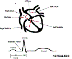
A schematic drawing of the normal heart structure and electric system (red arrows) is shown in Figure 1. A normal electric impulse originates in an area of the upper right atrium called the sinus (SA) node, and an ordinary electric activity of the heart is referred to as normal sinus rhythm. In normal sinus rhythm, an electric impulse is generated by the SA node, and that electricity spreads through the right and left atria, directing these chambers to beat.

On an electrocardiogram (ECG; bottom of Figure 1), the electric activity from the atria is seen as a small rounded deflection called a P wave. The same electric impulse then passes through a small area of tissue between the atria and ventricles called the atrioventricular (AV) node and then down through the ventricles. On the ECG, the electric activity from the ventricles results in a larger deflection called the R wave or QRS complex. Because the AV node is small, there is not enough electric activity to cause a deflection in the ECG. Therefore, the time spent by the electric impulse traveling through the AV node is represented on the ECG by a flat interval called the PR interval. In a normal heart, the AV node is a gatekeeper of sorts in that it is the only pathway for electricity that communicates from the upper chambers (atria) to the lower chambers (ventricles). The combination of electric impulses from the SA node to the atria, then through the AV node, and down to the ventricles results in 1 heartbeat. In summary, the electric activity on the ECG starts with a small deflection that results from atrial electric activity (the P wave), followed by a flat section resulting from electric activity through the AV node (the PR interval), followed by a larger deflection that results from electric activity from the ventricles (the R wave or QRS complex).


What Is an Accessory Pathway or Bypass Tract?

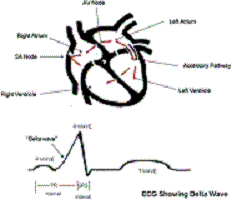
Some people are born with an extra piece of heart muscle tissue that connects directly between the atria and the ventricles, bypassing the AV node altogether. This abnormal piece of muscle is referred to as a bypass tract or an accessory pathway (Figure 2).

This extra piece of tissue can serve as a passageway for the electric signals between the atria and ventricles and allow electric activity in the ventricles to occur immediately after electric activity in the atria without having to wait for the electric impulse to travel through the AV node. In this situation, the ECG may not have much of a flat PR interval but instead may have the P wave (from atrial activity) right up against the R wave (from ventricular activity), with the R wave beginning with an upslope referred to as a delta wave (Figure 2). This delta wave results from electric activity traveling over the accessory pathway and bypassing the AV node.

Some people with an accessory pathway have a normal ECG at baseline because the accessory pathway is electrically active only when there is a fast, racing heartbeat (tachycardia) described below. Therefore, some people with accessory pathways can have completely normal ECGs like the one seen in Figure 1. This is referred to as a concealed accessory pathway.


How Does an Accessory Pathway Cause a Fast Heartbeat (Tachycardia)?

Not all patients with accessory pathways have fast heartbeats. Some people with a delta wave on their ECG caused by an accessory pathway will never have a problem with a fast heartbeat. In others, a rapid heartbeat ( tachycardia) can sometimes suddenly occur. The 3 different types of tachycardias associated with accessory pathways are shown in Figure 3.

The most common type (Figure 3 A) results from an electric circuit that travels from the atria through the AV node to the ventricles, then backward through an accessory pathway to the atria, and then back through the same circuit over and over again. Because this is a circuit that reenters itself and involves the atria and ventricles, it is referred to as AV reentrant tachycardia. The less common type of AV reentrant tachycardia (Figure 3 C) involves a circuit that travels forward from the atria through the accessory pathway and backward through the AV node.

Another type of tachycardia that can be seen in patients with accessory pathways is a chaotic irregular beating of the upper chambers of the heart called atrial fibrillation. If a patient with an accessory pathway develops atrial fibrillation and if that particular accessory pathway is capable of rapid electric conduction, this can lead to an extremely rapid pulse rate, which can be dangerous.

What is Wolff-Parkinson-White Syndrome?

Wolff-Parkinson-White syndrome (WPW) is the combination of accessory pathway activation seen on an ECG (delta waves) and episodes of tachycardia. It was first described in 1930 by Louis Wolff, Sir John Parkinson, and Paul Dudley White. Along with a delta wave, patients have a shorter time between the conduction of an impulse from the atrium to the ventricle, referred to as a short PR interval (see the ECG in Figure 2).

What is described above is called a preexcitation or WPW pattern. The presence of a pattern does not necessarily mean that the patient will experience WPW syndrome. The WPW pattern will be seen in about 0.2% of the general population. Of those patients with the WPW pattern, a minority will experience tachycardia and be defined as having WPW syndrome. WPW can be associated with Ebstein anomaly, a heart defect in which the valve connecting the right atrium and ventricle (tricuspid valve) is abnormally formed and placed lower than normal in the right ventricle. However, in most patients, WPW is not related to any other heart abnormality. It can occur at any age, is often first noted in childhood, but may not be diagnosed until adulthood in some patients. Symptoms of WPW syndrome are usually abrupt and may include palpitations, chest discomfort, and occasionally fainting.

On very rare occasions (less than 0.1% of the time), a patient with WPW can experience sudden cardiac death that results from the development of a chaotic irregular beating in the upper chambers of the heart called atrial fibrillation with rapid conduction down an accessory pathway (Figure 3B) leading to an extremely rapid pulse that can lead to cardiac arrest. Fortunately, this is a rare event in patients with WPW, and there are certain factors that a physician can often identify ahead of time to risk stratify patients with WPW.

Treatment

Treatment of WPW must be individualized. For patients who have only a documented WPW pattern on their ECG but no symptoms and no documented tachycardia, simple conservative observation may be appropriate. When symptoms or documentation of arrhythmias exists, a catheter-based electric evaluation of the heart called an electrophysiology study may be offered. This usually results in elimination of the accessory pathway with cautery delivered through the catheter called radiofrequency ablation. This will also eliminate the WPW pattern on the ECG. On rare occasions, WPW syndrome may be treated with antiarrhythmic medications, although most patients opt for the catheter ablation procedure because it may be curative and eliminate the need for lifelong medication therapy.

Conclusions

WPW syndrome results from tachycardia associated with an accessory pathway. The WPW pattern is diagnosed by a delta wave and/or short PR interval on an ECG. Once symptomatic, patients can be treated with catheter ablation or can try medications to control their symptoms. Management of WPW syndrome should be individualized, and treatment decisions should be carefully discussed with your doctor.

Reference: J.Kulig et al; Circulation 2010; 122: e480-e483.


Catecholaminergic Polymorphic Ventricular Tachycardia (CPVT)


What is Catecholaminergic Polymorphic Ventricular Tachycardia?

CPVT is a rare condition that affects the heart of otherwise fit and healthy people. It causes the heart to beat abnormally quickly, usually at times of exercise (particularly swimming) or times of high emotion, and can result in dizziness, sudden loss of consciousness or even death. It most commonly occurs in children and young adults typically in the first or second decade of life. It was first recognised in 1975.

In at least a third of cases, it is familial, (inherited) being passed down through the generations.

It is a difficult condition to diagnose, because all of the tests taken at a time of rest are normal including the electrocardiogram (ECG), and the echocardiogram (ultrasound of the heart).

The diagnosis is usually made by detecting extra beats, or runs of fast rhythm arising from the bottom part of the heart, (ventricles), during an exercise test or on a 24 hour ECG.

 How does CPVT get its name?

The heart is a pump that initiates its own electrical signals, causing it to beat between at between 50 and 180 beats per minute depending on whether you are resting, exercising or under some form of emotional/physical stress.

In response to stress the body releases adrenaline and noradrenaline (also called epinephrine and norepinephrine). Adrenaline and noradrenaline are chemical messengers that are collectively known as catecholamines that have a direct action on the heart. During exercise or emotional stimulation these catecholamines cause the heart to beat faster and the blood pressure to increase, thus enabling blood and oxygen to reach the exercising muscles and other vital organs.

This response to stress is how the body copes and allows us to flee from danger or run to catch the bus, however it also works the same if the body is emotionally or physically challenged in some other way, such as severe illness.

People with CPVT have hearts that respond abnormally to catecholamines, such that the heartbeat becomes rapid, chaotic and irregular at times of stress. A rapid heart rhythm is known as a “tachycardia”, and since this particular tachycardia arises from the main pumps or ventricles, it is known as a “ventricular tachycardia”. The word “polymorphic” literally means lots of different shapes- referring to the varying beats seen on the ECG. If such a rhythm sustains for more than a few seconds, insufficient blood reaches the brain, leading to a faint, collapse, or even death.

 How does it present?

CPVT typically presents with sudden collapse or severe dizziness, usually associated with exercise (or just after exercise stops) or intense emotional stress. First symptoms appear usually in the first or second decade of life, and may often be mistaken for epilepsy in children. It can also present quite out of the blue with sudden death.

 How can doctors make the diagnosis?

Symptoms may be reproduced by exercise test, and a 24-hour holter ECG (Holter) test can be useful. These tests are always performed in hospital under specialist supervision. Increasingly doctors use a long-term implanted digital loop recorder, or “Reveal” device, which records the ECG continuously, and documents the heart rhythm during a collapse.

Sudden collapse or severe dizziness may be associated with heart rhythm disturbance such as CPVT. Typically these  symptoms may occur with little or no warning, and are usually associated with exercise (or just after exercise stops) or   intense emotional stress. Typically cases present in the first or second decade of life, and may often be mistaken for epilepsy in children. Symptoms of the disease may be reproduced by exercise test or by using other forms of adrenergic stimulation such as drugs that increase the heartbeat. These tests are always performed in hospital under Specialist supervision.

Familial incidence of the condition has been manifest in 30% of cases to date.

 Treatment:

CPVT usually responds well to beta-blockers. Beta-blocker medications work to stop adrenaline and other catecholamines from speeding the heart up and therefore keep the heart rate under control. More recently, Flecainide has been found very effective in preventing this arrhythmia. In some cases beta-blockers are usually used in combination with implantable defibrillators (implantable cardioverter defibrillator “ICD”) that stop the fast chaotic heart rhythm by treating it with a small electric shock

 Genetic Diagnosis of CPVT:

At this time 3 genes are thought to be responsible for CPVT. Ryanodine (RYR2) mutations (abnormalities in the gene sequence), are thought to be responsible for the autosomal dominant (this means that each child born has a 50% chance of inheriting the mutation). In the heart ryanodine is located in the working part of the heart muscle cells that are responsible for making the heartbeat.

The role of ryanodine is to release calcium inside the cell in order for each heartbeat to occur, however this does not work properly as in the case of the abnormal ryanodine, then calcium may build up and not be realised as it should. When adrenaline is present and the heart rate speeds up the working cells are likely not able to cope with the increased workload and this throw the heart into the chaotic heart rhythm.

Calsequestrin CASQ2 binds to calcium inside the working areas of the cardiac muscle cells (sarcoplastic reticulum), it is thought that mutations in the calsequestrin prevent adequate binding occurring particularly when the cell is having to work hard in the presence of catecholamines such as adrenaline. The buildup of calcium because it cannot adequately attach itself to calsequestrin for transport is thought to cause the chaotic heart rhythm.

Ankyrin B mutations have also been implicated in CPVT, and these may also be linked to LQT (Long QT type 4). Work is still being done to establish this link.


Palpitations

Palpitations

Palpitations are a common problem that is often referred to a cardiologist for further investigation. Patients typically describe a sense that the heart is skipping or racing. This may be accompanied by shortness of breath, chest pain, and/or lightheadedness – or rarely, loss of consciousness. The sensation of palpitations can be caused by a wide variety of different things, ranging from normal heart behavior, to simple extra beats, all the way up to dangerous arrhythmias that could be life-threatening. The key goal of sorting this out is to record the heart rhythm at the time it is experienced. Typically, physicians will order an electrocardiogram (EKG or ECG) or a Holter monitor. Other tests your doctor may order include an echocardiogram, a stress test, or a coronary angiogram.

The trouble with ECGs and Holter monitors is that they are limited to a very short time. If the palpitations occur infrequently, these strategies will not obtain the desired symptom-rhythm correlation. Therefore, some patients will be asked to carry an Event Monitor, or a Loop Recorder – both monitors that can be worn for up to several weeks – in order to establish the diagnosis.

Once the nature of the palpitations is diagnosed, it can be treated appropriately.

Holter ECG


A special word about the most common cause of palpitations

One of the most common causes of palpitations is the skipping of the heartbeat that occurs from extra beats. These are simply extra beats that occur in all of us, but are felt more in some because they are more frequent. Most of the time these are simply beats from a secondary pacemaker in the heart that creates irregularity in the pulse that feels unusual. Typically, patients describe these as brief perturbations of the heart rhythm. The heart is otherwise normal, and the beats themselves are of no prognostic importance.

The heart has a single dominant pacemaker called the sinus node. There are several sites within the heart that are additional pacemakers that at times can be overactive. These sites can produce skipped beats that may occur as infrequently as 50 times a day, or as often as thousands of times a day. These do not normally represent anything serious, but can produce new symptoms when a sense of irregularity occurs. Patients will often describe the feeling of “heart jumping, flipping or missing”, with forceful beating after the skipped beat. This is illustrated in the adjacent figure, where the heart beat is regular followed by a skipped beat. After a short pause, there is a normal beat again. The pulse that this produces is illustrated below the trigger of the EKG and the figure. The normal pulse is followed by a very small pulse from the skipped beat. This is the sense of missing or skipping that occurs where the patient may feel the heart “stopping”. This is followed by a very forceful beat when in effect two heart beats are pumped out at once or the volume of two heart beats is pumped out all at once with the resumption of normal rhythm. This often leads to the sense that the heart skips or “misses count”, followed by a forceful pounding or impact in the chest with the normal beat.

This condition is benign, and is often an exaggerated version of the normal number of skipped beats that occurs in every person. Skipped beats are more common with any type of stimulant, including caffeine, decongestants, alcohol, bronchodilators (puffers for asthma) and occasionally with stress. Patients might find that modifying their lifestyle with respect to these agents reduces the frequency of skipped beats.

After recording these, reassurance is usually all that is necessary to confirm that there are no significant problems with the heart. Rarely, medication is used to suppress these skipped beats. Beta-blockers are used in the vast majority of patients when drug treatment is required. These agents reduce the adrenaline effect on the heart, making it less “irritable”. These are only used in patients who are very symptomatic, since there is no need to treat these beats for prognostic reasons.


** Source: Canadian Heart rhythm Society

Metabolic Syndrome

WHAT IS IT?

Metabolic syndrome is the name for a group of risk factors linked to overweight and obesity that increase your chance for heart disease and other health problems such as diabetes and stroke. The term “metabolic” refers to the biochemical processes involved in the body’s normal functioning.

Also Known As: Insulin Resistance Syndrome, Syndrome X

Basic Facts

  • Metabolic syndrome is the name for a group of risk factors that increase your chance for heart disease and other health problems such as diabetes and stroke.
  • The diagnosis of metabolic syndrome is made if you have any three out of these five heart disease risk factors: a large waistline, a higher than normal triglyceride level, a lower than normal level of HDL cholesterol (high-density lipoprotein cholesterol), higher than normal blood pressure, and higher than normal fasting blood sugar (glucose).
  • About 47 million adults in the United States (almost 25 percent) have metabolic syndrome. The rate of metabolic syndrome continues to rise as obesity levels increase.
  • Metabolic syndrome has several causes that act together. Some can be controlled, while others can’t.
  • To diagnose metabolic syndrome, your doctor evaluates your risk factors and checks your waist size, cholesterol, blood pressure, triglycerides, and blood sugar.
  • The first line of treatment for metabolic syndrome is lifestyle changes, including weight loss, increased physical activity, and a healthy diet. If lifestyle changes can’t control your risk factors, your doctor may recommend medicines.
  • Healthy lifestyle choices can help prevent metabolic syndrome. But if you develop metabolic syndrome, healthy lifestyle changes also can help to reverse or reduce the risk of heart disease and diabetes and complications of those conditions.
  • If you have metabolic syndrome, follow a healthy eating plan and increase your physical activity to manage your weight; quit smoking; and take all of your medicines as your doctor prescribes.

What Causes Metabolic Syndrome?

Metabolic syndrome has several causes that act together. Some can be controlled, while others can’t.

Causes that can be controlled include overweight and obesity, lack of physical activity, and insulin resistance.

Some causes you can’t control are growing older and genetics. Your chance of developing metabolic syndrome increases with age. Your genes can increase your chances of developing insulin resistance, for example, which can lead to metabolic syndrome, even if you have only a little extra weight around your waist.

Two other conditions are often found in people with metabolic syndrome, although it’s not known if they cause it or worsen it. The two conditions are a tendency to form blood clots and a tendency to have a constant, low-grade inflammation throughout the body.

Additional conditions that are being studied to see whether they have links to metabolic syndrome include:

  • Fatty liver (excess triglycerides and other fats in the liver)
  • Polycystic ovarian syndrome (a tendency to develop cysts on the ovaries)
  • Gallstones
  • Breathing problems during sleep such as sleep apnea

Who Is At Risk for Metabolic Syndrome?

You’re at greatest risk for metabolic syndrome if you have these underlying causes:

  • A large waistline (abdominal obesity)
  • Lack of physical activity
  • Insulin resistance

Some people are at risk for metabolic syndrome because the medicines they take may cause weight gain or changes in blood pressure, cholesterol, and blood sugar levels. These medicines are most often used for inflammation, allergies, HIV, and depression and other kinds of mental illnesses.

Populations Affected

About 47 million adults in the United States (almost 25 percent) have metabolic syndrome. Metabolic syndrome is more common in African American women than in African American men and in Mexican American women than in Mexican American men. It affects White women and men roughly equally.

Some racial and ethnic groups in the United States are more at risk for metabolic syndrome than others. Mexican Americans have the highest rate of metabolic syndrome (31.9 percent). Caucasians (23.8 percent) and African Americans (21.6 percent) have lower rates.

Other groups that are at increased risk of developing metabolic syndrome include:

  • People with a sibling or parent with diabetes
  • People with a personal history of diabetes
  • Women with a personal history of polycystic ovarian syndrome (a tendency to develop cysts on the ovaries)

In addition, members of certain ethnic groups are at increased risk for metabolic syndrome. For example, South Asians have an increased risk for metabolic syndrome.

Risk for Heart Disease

Having metabolic syndrome increases your risk for heart disease. Heart disease risk can be divided into short-term risk (the risk for having a heart attack or dying of heart disease in the next 10 years) and long-term risk (the risk for developing heart disease over your lifetime).

Other factors (aside from metabolic syndrome) contribute to your risk for heart disease as well. The major risk factors are:

  • Increased LDL cholesterol (low-density lipoprotein cholesterol) and total cholesterol levels. (LDL is the “bad” cholesterol.)
  • Cigarette smoking.
  • Blood pressure that is greater than or equal to 140/90 (or you’re on medicine for high blood pressure).
  • Decreased HDL cholesterol (high-density lipoprotein cholesterol) level to less than 40 mg/dL. (HDL is the “good” cholesterol.)
  • Age (for men ages 45 and older and for women ages 55 and older).
  • Family history of early heart disease or sudden death (in a father or brother before the age of 55, or in a mother or sister before the age of 65).

If you smoke cigarettes or have increased LDL cholesterol or high blood pressure, these are the first targets of treatment.

Regardless of whether you have metabolic syndrome, you should find out your chance of developing heart disease in the next 10 years (your short-term risk). This will help decide what your LDL cholesterol goal should be and how you should be treated. To start, you will need to count how many risk factors you have from the list above. (Don’t count LDL and total cholesterol because the treatment for them will be geared to your level of risk.)

  • You’re in the High Risk category for heart disease if you already have heart disease or diabetes, or if your 10-year-risk score is more than 20 percent.
  • You’re in the Moderately High Risk category if you have two or more risk factors and your 10-year-risk score is 10 to 20 percent.
  • You’re in the Moderate Risk category if you have two or more risk factors and your 10-year-risk score is less than 10 percent.
  • You’re in the Lower Risk category if you have zero or one risk factor.

Even if your 10-year-risk score isn’t high, over time metabolic syndrome will increase your chance for heart disease. This means that, regardless of your short-term risk category, metabolic syndrome should be treated (mainly with lifestyle changes).

What Are the Signs and Symptoms of Metabolic Syndrome?

Metabolic syndrome is made up of a group of factors that can increase risk even if they are only moderately raised (borderline-high risk factors). Metabolic syndrome itself usually has no symptoms. Most of the risk factors linked to metabolic syndrome have no signs or symptoms, although a large waistline is a visible sign.

Some people may have symptoms of high blood sugar (if diabetes is present) or, occasionally, high blood pressure. Symptoms of high blood sugar often include increased thirst; increased urination, especially at night; fatigue (tiredness); and blurred vision. High blood pressure is generally considered to have no signs or symptoms. However, a few people in the early stages of high blood pressure may have dull headaches, dizzy spells, or more nosebleeds than usual.

How Is Metabolic Syndrome Diagnosed?

The diagnosis of metabolic syndrome is based on the results of a physical exam and blood tests. To be diagnosed with metabolic syndrome, you must have at least three out of five of the following risk factors:

  • A large waistline. This means that you carry excess weight around your waist (abdominal obesity). Your doctor will measure your waist to determine whether you have abdominal obesity. A waist measurement of 35 inches or more for women and 40 inches or more for men is a component of metabolic syndrome and indicates an increased risk for heart disease and other health problems. A large waistline also is called “having an apple shape.”
  • A higher than normal triglyceride level, or you’re on medicine to treat high triglycerides. Triglycerides are a type of fat found in the blood. A triglyceride level of 150 mg/dL or higher is a component of metabolic syndrome.
  • A lower than normal level of HDL cholesterol (high-density lipoprotein cholesterol), or you’re on medicine to treat low HDL. HDL is considered “good” cholesterol because it lowers your chances of heart disease. An HDL cholesterol level less than 50 mg/dL for women and less than 40 mg/dL for men is a component of metabolic syndrome.
  • Higher than normal blood pressure, or you’re on medicine to treat high blood pressure. A blood pressure of 130/85 or higher is a component of metabolic syndrome. If only one of your two blood pressure numbers is high, it’s still a risk factor for metabolic syndrome.
  • Higher than normal fasting blood sugar (glucose), or you’re on medicine to treat high blood sugar. A normal fasting blood sugar is less than 100 mg/dL. Fasting blood sugar between 100 and 125 mg/dL is considered prediabetes. Fasting blood sugar of 126 mg/dL or higher is considered diabetes. A fasting blood sugar of 100 mg/dL or higher (prediabetes or diabetes) is a component of metabolic syndrome.

About 85 percent of people who have type 2 diabetes (the most common type) also have metabolic syndrome. These people have a much higher risk for heart disease than the 15 percent of people who have type 2 diabetes, but don’t have metabolic syndrome.

How Is Metabolic Syndrome Treated?

Healthy lifestyle changes are the first line of treatment for metabolic syndrome. Lifestyle changes include weight loss, increased physical activity, an improved diet, and quitting smoking.

Medicines are the next line of treatment. They’re used to treat and control individual risk factors such as high blood pressure, high triglycerides, low HDL cholesterol (high-density lipoprotein cholesterol), and high blood sugar. Medicines such as aspirin also may be used to reduce the risk of blood clots, a condition that often occurs with metabolic syndrome.

Goals of Treatment

The major goal of treating metabolic syndrome is to reduce a person’s risk for heart disease. Treatment is directed first at reducing LDL cholesterol (low-density lipoprotein cholesterol), high blood pressure, and diabetes (if these conditions are present).

The second goal of treatment is to prevent the onset of type 2 diabetes (if it hasn’t already developed). Long-term complications of diabetes often include heart and kidney disease, vision loss, and foot or leg amputation. If diabetes is present, the goal of treatment is to reduce the increased risk for heart disease by controlling all of the risk factors.

The main emphasis in the treatment of metabolic syndrome is to lessen the effects of the underlying risk factors that can be controlled, such as overweight, lack of physical activity, and an unhealthy diet.

Specific Types of Treatment

Weight Loss

In general, people with metabolic syndrome who are overweight or obese are urged to reduce their weight by 7 to 10 percent during the first year of treatment. For example, a person weighing 250 pounds should try to lose 18 to 25 pounds. A person weighing 300 pounds should try to lose 21 to 30 pounds.

After the first year, people are urged to continue to lose weight to the extent possible, with a long-range target of lowering their body mass index (BMI) to less than 25. BMI measures your weight in relation to your height and gives an estimate of your total body fat. A BMI between 25 and 29.9 is considered overweight. A BMI of 30 or more is considered obese. A BMI of less than 25 is the goal for prevention and treatment of metabolic syndrome.

Healthy Eating Plan

With the TLC diet, less than 7 percent of your daily calories should come from saturated fat, and no more than 25 to 35 percent of your daily calories should come from all fats, including saturated, trans, monounsaturated, and polyunsaturated fats. You also should consume less than 200 mg a day of cholesterol. The amounts of fat and cholesterol in prepared foods can be found on the food’s nutritional label.

Foods high in soluble fiber also are part of a healthy eating plan. These foods include:

  • Whole grain cereals such as oatmeal and oat bran
  • Fruits such as apples, bananas, oranges, pears, and prunes
  • Legumes such as kidney beans, lentils, chick peas, black-eyed peas, and lima beans

Fish are an important part of a heart healthy diet. Fish are a good source of omega-3 fatty acids, which may help protect the heart from blood clots and inflammation and reduce the risk for heart attack.

You also should try to limit the amount of sodium and salt that you eat. This means choosing low-sodium and low-salt foods and “no added salt” foods and seasonings at the table or when cooking. The nutritional label on food packaging shows the amount of sodium in the item.

Try to limit alcoholic beverages. Too much alcohol will raise your blood pressure and triglyceride level. It will also add extra calories, which will cause weight gain. Men should have no more than two drinks containing alcohol a day. Women should have no more than one drink containing alcohol a day.

Increased Physical Activity

In general, people with metabolic syndrome are urged to keep up a moderate level of activity, such as brisk walking for at least 30 minutes at least 5 days of the week. This activity can be broken into shorter periods as needed—for example, three 10-minutes sessions.

The ultimate goal is for people to maintain a moderate level of physical activity 60 minutes a day for 5 days a week, but preferably daily. You should talk with your doctor about the best kind of physical activity for you before starting any kind of program.

Smoking

If you smoke, quitting is important. Among other known harmful effects on your heart, smoking will raise your triglyceride level and lower your HDL cholesterol.

Medicines

Your doctor may recommend medicines to help treat unhealthy cholesterol levels, high blood pressure, and high blood sugar. Unhealthy cholesterol levels are treated by one or more cholesterol-lowering medicines such as statins, fibrates, or nicotinic acid.

High blood pressure is treated by one or more antihypertensive medicines such as diuretics or angiotensin-converting enzyme (ACE) inhibitors. High blood sugar is treated with oral medicines (such as metformin), insulin injections, or both. Low-dose aspirin can help reduce the risk of forming blood clots, especially for people at high risk for heart disease.

How Can Metabolic Syndrome Be Prevented?

Making healthy lifestyle choices is the best way to prevent metabolic syndrome. Maintaining a healthy weight is important. Other than weighing yourself on a scale, there are two ways to know whether you’re at a healthy weight: waist measurement and body mass index (BMI).

A waist measurement indicates your abdominal fat and is linked to your risk for heart disease and other diseases. To measure your waist, stand and place a tape measure around your middle, just above your hipbones. Measure your waist just after you breathe out. Make sure the tape is snug but doesn’t squeeze the flesh. A waist measurement of less than 35 inches for women and less than 40 inches for men is the goal for preventing metabolic syndrome; it’s also the goal when treating metabolic syndrome.

BMI measures your weight in relation to your height and provides an estimate of your total body fat. A BMI between 25 and 29.9 is considered overweight. A BMI of 30 or more is considered obese. A BMI of less than 25 is the goal for preventing metabolic syndrome, and it’s also the goal when treating metabolic syndrome.

To maintain a healthy weight, follow a healthy eating plan and try not to overeat. This means eating fewer calories and less saturated fat, and emphasizing whole grains, fish, and fruits and vegetables. Choose unsaturated fats when eating fats and oils such as canola, olive, or safflower oils, soft or liquid margarine, and nuts.

Increasing your physical activity also can help you maintain a healthy weight. Talk to your doctor about what kind of physical activity is best for you. If you’re medically able, get at least 30 minutes of moderate activity, such as brisk walking, at least 5 days a week. With your doctor’s permission, work up to getting 60 minutes of moderate activity 5 to 7 days a week.

Make sure to schedule regular doctor visits to keep track of your cholesterol, blood pressure, and blood sugar levels. A cholesterol blood test will show your levels of LDL cholesterol (low-density lipoprotein cholesterol), HDL cholesterol (high-density lipoprotein), and triglycerides.

Living With Metabolic Syndrome

Metabolic syndrome is a lifelong condition. However, lifestyle changes can help you reverse or reduce your risk factors and reduce your risk of heart disease and diabetes. If you already have heart disease and/or diabetes, lifestyle changes can help you prevent or delay complications such as heart attack, stroke, and the long-term effects of diabetes (damage to your eyes, nerves, kidneys, feet, and legs).

Making healthy lifestyle changes can include following a healthy eating plan, increasing your physical activity, and quitting smoking. Part of managing metabolic syndrome also includes taking all of your medicines as your doctor prescribes.

Make realistic short- and long-term goals for yourself when you begin to make healthy lifestyle changes. Work closely with your doctor and seek regular medical care.


**Source: Cardiosource- American College of Cardiology.