What Is a Normal ECG in Athletes?

Up to 60% of athletes show ECG changes, and some of these changes are often striking to look at.  The Seattle ECG criteria is often used to separate the normal from the abnormal ECG in an athlete. This is important, as many normal athletes undergo cardiac angiography and even pacemaker surgery because of misinterpretation of their ECGs.

  • Bradycardia: A low heart rate in an athlete should be self-evident—but it is not in the real world. Common normal athletic ECG patterns include: the inverted P-wave and junctional rhythm, Mobitz type I Wenckebach block, and sinus arrhythmia.
  • Incomplete right bundle branch block (IRBBB): While only 10% of the general population has IRBBB, up to 40% of athletes have it as a normal pattern. This might be due to remodeling of the athletic right ventricle.
  • Left ventricular hypertrophy (LVH): It is normal for athletes to have voltage criteria for LVH. Of course, it is abnormal when voltage criteria comes with other LVH related patterns, such as left atrial abnormality, ST-segment strain patterns, left axis deviation, etc.
  • Early repolarization: It is common and normal for athletes to have early repolarization, including terminal QRS notching. The pattern is especially common in black athletes.
  • T-wave changes: T-wave inversions can be normal, or they can be abnormal. Up to one in three black athletes have terminal T-wave inversions in leads V1–V4. T-wave inversion patterns which can be mistaken for anterior-wall ischemia. Paying attention to the location of the inversions, however, is important. Namely, it is normal to have T-wave inversions in leads V1–V4 but not in V5 or V6 and not with accompanying QRS abnormalities or frequent premature ventricular contractions (PVCs).
  • Notable abnormal findings in athletes: Some of the abnormal ECG findings may include any complete left bundle branch block, intraventricular conduction delay (IVCD) greater than 140 ms, left atrial abnormality, right ventricular hypertrophy, Wolff-Parkinson-White (WPW) syndrome, QT prolongation (greater than 470 ms in males and 480 ms in females), high-degree AV block, atrial fibrillation/flutter, and high-density PVCs. Isolated (low-density) PVCs are considered benign.
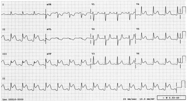
 

55-year old male with chest pain.

The following ECG was taken from a  55-year old male who presented to the ER with 2-hour history of persistent heavy discomfort across the anterior chest with radiation to the neck.

Risk factors: Positive family history (father had MI at age 52); hypertension; dyslipidemia.

Medications: Nifedipine XL 30 mg daily and atorvastatin 10 mg daily.

Physical examination: Heart rate 100 beats per minute, BP 130/85 mmHg,  respiratory rate 22/minute. Jugular veinous pressure is 4 cm above the sternal angle at 45 degrees. Soft S1 and S2 and prominent S4. No murmurs.

Questions:

1)- What is the ECG diagnosis?

a. Acute pericarditis.

b. Acute anterior ST elevation myocardial infarction.

c. Acute infero-posterior-lateral ST elevation myocardial infarction.

d. Acute infero-posterior-lateral with right ventricular myocardial infarction.

New guidance for management of asymptomatic young people with Wolff-Parkinson-White (WPW) syndrome

A consensus statement on the management of asymptomatic patients with Wolff-Parkinson-White (WPW) syndrome has been presented at The Heart Rhythm Society (HRS) 2012 Scientific Sessions. This statement should help clarify which young people should undergo catheter ablation. While there is a small chance that an asymptomatic young person could end up having a life-threatening arrhythmia, the number is not zero. Yet, catheter ablation for every young individual who has ever had a WPW pattern on his/her electrocardiogram (ECG) is also not the answer.

Cohen and colleagues released the statement on May 18, 2012 at the Heart Rhythm Society Scientific Sessions.

The guidance is explicitly directed at physicians treating young patients with WPW and defines young people as between eight and 21. At the crux of the document is the question of just when physicians should intervene—and when they shouldn’t—in young people found, on ECGs, to have the signature electrocardiographic WPW pattern. These are increasingly important questions, given increased emphasis on pre-participation screeningfor sports in young people.

The expert consensus statement, a joint effort of the Pediatric and Congenital Electrophysiology Society (PACES) and the HRS, estimates that from one to three young people per 1000 likely have WPW, although many—around 65%—are asymptomatic.

The guideline writers recommend the following:

  • If a child is old enough to comply, an exercise stress test to look for persistent preexcitation is “reasonable.” Clear loss of preexcitation at physiologic heart rates is associated with lower risk of sudden death due to accessory pathways.
  • Where noninvasive testing shows persistent or uncertain loss of preexcitation, diagnostic transesophageal or intracardiac electrophysiology studies are warranted. Recommendations, based on test results, include ablation or continued awareness and observation for symptoms.
  • Ablation is a “reasonable” consideration in young people with a shortest preexcited RR interval (SPERRI) <250 ms. Young people with SPERRI >250 ms are lower risk, and ablation may be deferred.
  • Patients deemed low risk who subsequently develop symptoms like syncope or palpitations may be eligible for ablation.
  • WPW in the setting of structural heart disease increases risk for both atrial tachycardia and atrioventricular (AV) reciprocating tachycardia. Patients can be considered for ablation.
  • Ablation may also be considered in asymptomatic patients with WPW who have ventricular dysfunction secondary to dyssynchronous contractions.


Source: The heart.org


Subscribe to Our Newsletter

Lorem ipsum dolor amet consetetur sadipscing elitr diam nonumy eirmod tempor invidunt ut.
© All rights reserved.
Powered by YOOtheme.