Valve Prosthesis–Patient Mismatch

The concept/phenomenon of valve prosthesis/patient mismatch (VP–PM) was first described in 1978.  All prosthetic heart valves have some degree of VP–PM.

The original paper (in 1978) that described valve prosthesis–patient mismatch (VP–PM) stated that “Mismatch can be considered to be present when the effective prosthetic heart valve area, after insertion into the patient, is less than that of a normal human valve”. It must be noted that all prosthetic heart valves (PHVs) are smaller than normal and thus are inherently stenotic.  The PHV mismatch was stated to be “usually mild to moderate in severity and often of no immediate clinical significance.” However, severe mismatch may lead to significant symptomatic and hemodynamic deterioration and increased mortality.

With the publication of VP–PM, surgeons were more careful and inserted the largest PHV that could be safely inserted. PHVs with improved hemodynamic profile have also been developed. As a result severe VP–PM has become a much less common clinical problem. The overwhelming majority of a large number of scientific publications have related to the aortic valve and not to mitral valve VP–PM.

How should VP–PM be measured?

Different parameters have been evaluated to assess for the severity of VP–PM. The most commonly used measures of valve size is the EOAi.

The EOA is a physiological parameter analogous to the native AVA. The EOA can be measured both invasively and noninvasively using echocardiography/Doppler or magnetic resonance imaging. The most readily and widely available method is echocardiography/Doppler. The accuracy of EOA echocardiographic measurement in the bioprosthetic valve is limited by the same pitfalls that are present in the measurement of the AVA; in particular, the LV outflow tract diameter may be more difficult to measure because of reverberation artifact caused by the prosthetic heart valve, but in these instances, the sewing ring diameter may be a sufficient surrogate. In the bileaflet mechanical valve, the central orifice may produce a high-velocity jet, causing an underestimation of the EOA. Pressure recovery occurs with both bioprosthetic and mechanical heart valves, although the implications of pressure recovery in PHVs have not yet been clarified. The EOAi has a complex relationship with the mean gradient across the aortic valve and PHV.

Assessment of severity of VP–PM

With aortic VP–PM the obstruction to the LV outflow tract is similar to that seen with native AS. Thus, the severity of aortic VP–PM should be assessed by the same criteria as for severe AS. Severe aortic stenosis is defined as mean aortic valve gradient, measured after energy recovery (also called pressure recovery), of ≥50 mm Hg and an AVA index of ≤ 0.6 cm²/m². ); it would be reasonable that this criterion should also be applied to severe VP–PM.


Grading

AVA and EOA, cm2

AVA Index and EOAi, cm2/m2

Mild

>1.5

>0.9

Moderate

>1.0–1.5

>0.6–0.9

Severe

≤1.0

≤0.6

Very severe/critical

≤0.7

≤0.4


When should the severity of VP–PM be determined?

4 phases of physiological healing of mechanical and bioprosthetic PHVs have been described; they are “platelet and fibrin deposition, inflammation, granulation tissue, and finally encapsulation. Long-term device fibrous encapsulation with extension to adjacent tissues adds to structural stability.” Bioprosthetic valves undergo morphological changes of both the tissue material as well as the supporting structures, which may contribute to VP–PM. Valve leaflets become covered by fibrin, platelets, and other cellular material. The matrix of the leaflets undergoes microcalcification as well insulation with plasma materials, causing changes in the matrix structure. These changes may change the resistive properties of leaflet materials. In both mechanical and bioprosthetic valves, a fibrous sheath may also encapsulate the supporting structure of the valve, encroaching on the PHV orifice and also possibly causing valve leaflet or disk immobilization.

For PHVs of the same size, there was a wide range of EOAs. Two echocardiographic/Doppler studies from the Mayo Clinic of patients studied within 1 week of mitral porcine mitral bioprosthesis and of tricuspid mechanical prostheses showed a wide range of gradients and EOAs with the same size of PHV, even in the same brand of PHV.

There are at least several explanations for these findings. 1) PHVs of the same labeled size are not necessarily of precisely the same size. Bioprostheses and other biological valves may also have differences in tissue materials. 2) There are variations from patient to patient with regard to healing changes, hemodynamic conditions, and pressure recovery.

It is best to measure VP–PM early (at 1 week after PHV implantation or at time of hospital discharge) to determine the variations in actual size of the PHV that was implanted in an individual patient. Importantly, VP–PM should also be assessed at 6 to 12 months when the physiological and other morphological changes in the PHV are mostly complete. The severity of VP–PM determined at this time can be expected to determine the long-term impact of VP–PM on patients’ outcomes.

Conclusions:

It must be noted that all prosthetic heart valves (PHVs) are smaller than normal and thus are inherently stenotic.

1) – EOAi should be measured at 1 to 4 weeks or at hospital discharge to evaluate the actual valve size that was implanted. This should also be done at 6 to 12 months to evaluate the severity of VP–PM that will affect long-term outcomes.

2) – The grading of severity of VP–PM should be similar to another common LV outflow tract obstruction, namely, valvular AS. VP–PM can be mild (EOAi >0.9 cm2/m2), moderate (EOAi >0.6 to 0.9 cm2/m2), or severe (EOAi ≤0.6 cm2/m2).

3) – Mild VP–PM, like mild AS, is unlikely to have clinically significant untoward effects. Outcomes with moderate and severe VP–PM should be evaluated separately. Moderate VP–PM is unlikely to reduce survival unless there is progression of valve obstruction, for example, with pannus formation. Severe VP–PM has negative effects on outcomes, but its effect on mortality is still unproven.

4) – Prediction of severity of VP–PM is problematic. The primary goal should be not to prevent VP–PM but rather to prevent severe VP–PM.

5) – Use of the EOAi as a continuous variable may help to define the level of severe VP–PM that results in increased mortality, and this may occur at a critical level of obstruction (≤0.4 cm2/m2).
 

References:
1)- Rahimtoola S, The problem of valve prosthesis-patient mismatch, Circulation 1978; 58:20-24.                                                                                                                                                                                                                                                                                                                  2)- Daneshuar S.A.; Rahimtoola S.H. Valve Prosthesis-Patient Mismatch (VP-PM): Long-Term Prespective. J AM Coll Cardiol 2012;60(13):1123-1135.

Wolff-Parkinson-White Syndrome

The heart has 4 chambers that pump blood. The 2 upper chambers are the right and left atria, and the 2 lower chambers are the right and left ventricles. The heart also has an electric system that directs the coordinated beating of these 4 chambers.

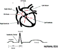
A schematic drawing of the normal heart structure and electric system (red arrows) is shown in Figure 1. A normal electric impulse originates in an area of the upper right atrium called the sinus (SA) node, and an ordinary electric activity of the heart is referred to as normal sinus rhythm. In normal sinus rhythm, an electric impulse is generated by the SA node, and that electricity spreads through the right and left atria, directing these chambers to beat.

On an electrocardiogram (ECG; bottom of Figure 1), the electric activity from the atria is seen as a small rounded deflection called a P wave. The same electric impulse then passes through a small area of tissue between the atria and ventricles called the atrioventricular (AV) node and then down through the ventricles. On the ECG, the electric activity from the ventricles results in a larger deflection called the R wave or QRS complex. Because the AV node is small, there is not enough electric activity to cause a deflection in the ECG. Therefore, the time spent by the electric impulse traveling through the AV node is represented on the ECG by a flat interval called the PR interval. In a normal heart, the AV node is a gatekeeper of sorts in that it is the only pathway for electricity that communicates from the upper chambers (atria) to the lower chambers (ventricles). The combination of electric impulses from the SA node to the atria, then through the AV node, and down to the ventricles results in 1 heartbeat. In summary, the electric activity on the ECG starts with a small deflection that results from atrial electric activity (the P wave), followed by a flat section resulting from electric activity through the AV node (the PR interval), followed by a larger deflection that results from electric activity from the ventricles (the R wave or QRS complex).


What Is an Accessory Pathway or Bypass Tract?

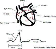
Some people are born with an extra piece of heart muscle tissue that connects directly between the atria and the ventricles, bypassing the AV node altogether. This abnormal piece of muscle is referred to as a bypass tract or an accessory pathway (Figure 2).

This extra piece of tissue can serve as a passageway for the electric signals between the atria and ventricles and allow electric activity in the ventricles to occur immediately after electric activity in the atria without having to wait for the electric impulse to travel through the AV node. In this situation, the ECG may not have much of a flat PR interval but instead may have the P wave (from atrial activity) right up against the R wave (from ventricular activity), with the R wave beginning with an upslope referred to as a delta wave (Figure 2). This delta wave results from electric activity traveling over the accessory pathway and bypassing the AV node.

Some people with an accessory pathway have a normal ECG at baseline because the accessory pathway is electrically active only when there is a fast, racing heartbeat (tachycardia) described below. Therefore, some people with accessory pathways can have completely normal ECGs like the one seen in Figure 1. This is referred to as a concealed accessory pathway.


How Does an Accessory Pathway Cause a Fast Heartbeat (Tachycardia)?

Not all patients with accessory pathways have fast heartbeats. Some people with a delta wave on their ECG caused by an accessory pathway will never have a problem with a fast heartbeat. In others, a rapid heartbeat ( tachycardia) can sometimes suddenly occur. The 3 different types of tachycardias associated with accessory pathways are shown in Figure 3.

The most common type (Figure 3 A) results from an electric circuit that travels from the atria through the AV node to the ventricles, then backward through an accessory pathway to the atria, and then back through the same circuit over and over again. Because this is a circuit that reenters itself and involves the atria and ventricles, it is referred to as AV reentrant tachycardia. The less common type of AV reentrant tachycardia (Figure 3 C) involves a circuit that travels forward from the atria through the accessory pathway and backward through the AV node.

Another type of tachycardia that can be seen in patients with accessory pathways is a chaotic irregular beating of the upper chambers of the heart called atrial fibrillation. If a patient with an accessory pathway develops atrial fibrillation and if that particular accessory pathway is capable of rapid electric conduction, this can lead to an extremely rapid pulse rate, which can be dangerous.

What is Wolff-Parkinson-White Syndrome?

Wolff-Parkinson-White syndrome (WPW) is the combination of accessory pathway activation seen on an ECG (delta waves) and episodes of tachycardia. It was first described in 1930 by Louis Wolff, Sir John Parkinson, and Paul Dudley White. Along with a delta wave, patients have a shorter time between the conduction of an impulse from the atrium to the ventricle, referred to as a short PR interval (see the ECG in Figure 2).

What is described above is called a preexcitation or WPW pattern. The presence of a pattern does not necessarily mean that the patient will experience WPW syndrome. The WPW pattern will be seen in about 0.2% of the general population. Of those patients with the WPW pattern, a minority will experience tachycardia and be defined as having WPW syndrome. WPW can be associated with Ebstein anomaly, a heart defect in which the valve connecting the right atrium and ventricle (tricuspid valve) is abnormally formed and placed lower than normal in the right ventricle. However, in most patients, WPW is not related to any other heart abnormality. It can occur at any age, is often first noted in childhood, but may not be diagnosed until adulthood in some patients. Symptoms of WPW syndrome are usually abrupt and may include palpitations, chest discomfort, and occasionally fainting.

On very rare occasions (less than 0.1% of the time), a patient with WPW can experience sudden cardiac death that results from the development of a chaotic irregular beating in the upper chambers of the heart called atrial fibrillation with rapid conduction down an accessory pathway (Figure 3B) leading to an extremely rapid pulse that can lead to cardiac arrest. Fortunately, this is a rare event in patients with WPW, and there are certain factors that a physician can often identify ahead of time to risk stratify patients with WPW.

Treatment

Treatment of WPW must be individualized. For patients who have only a documented WPW pattern on their ECG but no symptoms and no documented tachycardia, simple conservative observation may be appropriate. When symptoms or documentation of arrhythmias exists, a catheter-based electric evaluation of the heart called an electrophysiology study may be offered. This usually results in elimination of the accessory pathway with cautery delivered through the catheter called radiofrequency ablation. This will also eliminate the WPW pattern on the ECG. On rare occasions, WPW syndrome may be treated with antiarrhythmic medications, although most patients opt for the catheter ablation procedure because it may be curative and eliminate the need for lifelong medication therapy.

Conclusions

WPW syndrome results from tachycardia associated with an accessory pathway. The WPW pattern is diagnosed by a delta wave and/or short PR interval on an ECG. Once symptomatic, patients can be treated with catheter ablation or can try medications to control their symptoms. Management of WPW syndrome should be individualized, and treatment decisions should be carefully discussed with your doctor.

Reference: J.Kulig et al; Circulation 2010; 122: e480-e483.


Questions if you are considering a heart surgery!

What to ask your cardiologist…

  • What   is coronary artery disease? How many heart vessels are blocked?

  • What   are my treatment options and what are the risks and benefits of each?

  • Why   are you recommending this treatment over the others?

  • What   lifestyle changes will I need to make and what community resources are   available to help?

  • If   I decide not to have open-heart surgery, will you support my decision?

  • Will   I be given some time to put my affairs in order prior to the heart surgery?

  • Should   I complete an Advance Directive?

  • Should   I avoid caffeine and other stimulants for 48 hours prior to surgery?

  • (If   diabetic) How often should I be monitoring my blood sugar?

  • If   I opt to have surgery, will I need to take medication afterwards? If so, for   how long?

What to ask your heart surgeon…

  • What   are the risks and complications associated with this surgery?

  • What   are my specific risks? How risky is my surgery?

  • Did   I have a heart attack? If so, how will this affect my surgery?

  • How   many other (men / women) have you operated on with my same condition and what   were their outcomes?

  • How   many patients do you operate on each year? How many of your patients were   women?

  • Can   my chest and leg scars be minimized or can vessels be taken from a less   visible location?

  • How   long is my expected recovery? When will I be able to resume normal   activities? Drive a car? Return to work? Have sex? Be independent in   activities of daily living? Make plans for help in the home for the first   full two weeks following your discharge from the hospital. Ask for a social   service referral to help assist your family with sharing duties.

  • What   is your fee? Will you accept my insurance as payment in full?

  • If   I decide not to have blood products administered, does the hospital follow   Bloodless Care Protocols?

  • What   accommodations are available for my family’s stay on the hospital campus so   that they can support me throughout the operation?

  • When   will you be available to answer some of my concerns about the operation? Write   down your appointment time to speak with your surgeon or the cardiac team   nurse:Date _________ Time__________

Tell your heart surgeon…

  • If   you want bloodless care;

  • If   you want your Advance Directives postponed during surgery. This is the usual   procedure. It will be reinstated after you are stable and transferred to   intensive care;

  • If   you have had a vein stripping operation. The doctor will need to look   elsewhere for vessels;

  • If   you would like to hear reassurances during the operation that “all is   going well”;

  • If   you would like to employ holistic measures to help you relax, for example   therapeutic massage, aroma therapy, reflexology, bio-feedback or music   therapy.

  • If   you would like to play a relaxing CD during the surgery. You may need to   bring in your own headphones and CD player, but this may be well worth the   trouble.

Discuss with your anesthesiologist any health history that could   affect how you respond to anesthesia, for example, if you…

  • Have   panic attacks

  • Experience   chest pains not related to activity

  • Take   medication for anxiety

  • Excessively   drink, binge drink or do weekend drinking

  • Take   herbal supplements

  • If   you or a family member (blood relative) has ever had a serious reaction to   anesthesia;

  • If   you have a drug allergy or sensitivity or you are allergic to latex.

List drug allergies here:
 
 
_____________________________________________________

Source: Women’s Health Foundation

Hypertrophic Cardiomyopathy (HCM)

Hypertrophic cardiomyopathy is a condition where the heart muscle becomes thickened. The symptoms that develop depend on the severity of the condition. The treatment depends on the type of symptoms you have and whether complications develop. Some people need no treatment. Most cases are hereditary so screening of close family members is advised.

What happens in hypertrophic cardiomyopathy?

In HCM the heart muscle becomes thickened (hypertrophies) in parts of the heart. In the normal heart, the muscle cells are regular and patterned. In HCM the cells of the heart muscle become irregular and disordered.

The muscle surrounding the left ventricle is the area commonly affected. Sometimes the muscle around the right ventricle is also affected. The degree of thickening may vary in different places. For example, the septum (the wall dividing the right and left ventricle) is often the area with the greatest thickening. In about 1 in 4 people the muscle thickening is evenly distributed throughout the walls of the left ventricle.

The thickened heart muscle usually contracts well to pump blood out of the heart. However, it may lead to problems which include the following:

  • The affected heart muscle (usually around the left ventricle) may become stiff. This can mean that your left ventricle may not fill as  easily as normal. Less blood than normal is then pumped out from your heart with each heartbeat.
  • The thickening is often most marked in the upper part of the septum. This may partly obstruct the flow of blood from your left  ventricle into your aorta. This results in less blood being pumped out from your heart.
  • The thickened heart muscle may affect the function of your heart  valves. In particular, the mitral valve may become leaky if it does not close properly.
  • In some people, the abnormal heart muscle affects the electrical conducting system of the heart. This may cause abnormal heart rates and/or rhythms to develop.

What causes hypertrophic cardiomyopathy?

Heart muscle can thicken because of something, such as high blood pressure. In HCM the heart muscle thickens without an obvious cause.

In most cases the condition is inherited. If a couple (where one person has HCM) has a child, there is a 1 in 2 chance of the child being affected. This pattern of inheritance is called autosomal dominant. It seems that affected people inherit defective genes which are involved in making parts of the heart muscle cells.

Men and women are affected equally.

Who develops hypertrophic cardiomyopathy?

It is sometimes present at birth and can develop in young children. However, it most commonly develops in the teenage years or early adulthood.

What are the symptoms of hypertrophic cardiomyopathy?

If you only have mild thickening of the heart muscle you may not have any symptoms. Symptoms can range from mild to severe and may not develop straight away. Possible symptoms include the following:

  • Shortness of breath. This may develop only when you exercise if the condition is mild. When the condition is more severe, you can be breathless at rest.
  • Chest pain (angina). This may develop only when you exercise, but can also occur at rest when it is more severe. The pain occurs because the supply of blood and oxygen to the heart muscle is not sufficient to meet the demands of the thickened muscle.
  • Palpitations. Sometimes abnormalities of heart rhythm (arrhythmias) develop which can cause palpitations. You      may become aware that your heartbeat is fast and/or irregular.
  • Dizziness and fainting attacks. These occur more commonly when you exercise, but may occur when you are resting. This may be due to reduced output of blood from the heart or because of arrhythmias.

How is hypertrophic cardiomyopathy diagnosed?

A doctor may suspect this condition because of:

  • Your symptoms.
  • Your family history.
  • Changes on your electrocardiogram (ECG) – this is a tracing of the electrical activity of the heart.
  • Changes on your chest X-ray. This may show your heart is large or that there is fluid in your lungs.
  • Ultrasounds scan of the heart. This is called an echocardiogram.   This is a painless test which can measure the thickness of your heart      muscle.

Once the diagnosis is confirmed, other tests may be needed to assess the severity of your condition. A Doppler ultrasound scan also looks at blood flow through the heart chambers. This shows how well the heart ventricles are filling and contracting. A Doppler ultrasound scan can also show if there is any turbulent blood flow within the ventricles.

How does the condition progress?

The thickening of the heart muscle does not tend to progress once you stop growing. This means that, for many people, the symptoms remain stable during adulthood. Unfortunately, the symptoms gradually become worse for some people as the heart muscle becomes stiffer. Sometimes the function of the heart gradually deteriorates and heart failure may develop.

Complications may occur and include the following:

Arrhythmias

An arrhythmia is an abnormal rate or rhythm of the heartbeat. There are various types of arrhythmia, some more serious than others. Sometimes an arrhythmia develops intermittently and can cause bouts of palpitations, dizziness and other symptoms. Some arrhythmias become permanent. Arrhythmias can usually be treated.

Endocarditis

This is a rare complication. Endocarditis is an infection of the inside lining of the heart chambers and heart valves. Unless promptly treated, endocarditis can cause serious illness.

People with HCM used to be advised to take antibiotics before dental treatment or other procedures. This is no longer the case, as taking antibiotics has not been shown to reduce the risk of developing infective endocarditis.

Sudden death

Sudden collapse and death occurs in a small number of people with HCM. This is probably due to a severe arrhythmia which may develop suddenly. People most at risk are those with more severe disease, especially those who have had a previous serious arrhythmia. Some people at high risk may be advised on treatments which aim to prevent or treat arrhythmias.

What is the treatment for hypertrophic cardiomyopathy?

There is no treatment which can reverse the changes of the heart muscle. Treatment aims to ease symptoms if they occur and to prevent complications. If you do not have any symptoms or you only have mild symptoms then you may not need any treatment. Treatment which may be required includes the following:

Medication

The medicines advised depend on what symptoms or complications develop. For example:

  • Beta-blockers (such as propranolol)      and calcium antagonists (especially verapamil) are the commonly used  medicines. These can slow the heart rate and make the heart contract less  forcefully. This allows more time for the ventricle to fill with each  heartbeat. These medicines may be used to treat chest pain, breathlessness and palpitations.
  • Various other medicines called anti-arrhythmic medicines (for example, amiodarone) are used to treat and to prevent arrhythmias.   They work by interfering with and helping to correct the electrical  impulses in your heart.
  • Warfarin may be advised if you  develop atrial fibrillation (a common arrhythmia). With this arrhythmia a blood clot is a possible complication. Warfarin is an anticoagulant. This means it helps to prevent blood clotting by thinning the blood.

Other types of treatment for arrhythmias

Other treatments may be an option if you develop arrhythmias. For example:

  • Cardioversion is an option for some types of arrhythmia. Whilst under anaesthetic, you are given an electric shock over the heart. This may revert the rhythm back to normal.
  • Artificial pacemakers are used in certain types of arrhythmia where the heart beats abnormally slowly (complete heart block) and in certain other situations. An artificial pacemaker is a small device which is inserted just under the skin on the upper chest.  Wires from the pacemaker are passed through veins into the heart chambers.  The pacemaker can then stimulate the heart to maintain a regular normal heartbeat.
  • Implantable cardioverter defibrillators (ICDs) are sometimes used in certain situations – especially if you are at risk of developing serious and life-threatening arrhythmias. They are small devices which are similar to pacemakers and are inserted under the  skin in the upper chest. Wires are passed through a vein to the heart. The device monitors the heartbeat. If it detects an abnormal rhythm, the device can send a small electrical shock to the heart to change it back to normal.

Surgery

If your cardiomyopathy is severe, an operation may be option:

  • Myectomy. This is an operation to remove a segment of thickened muscle from the septum. It is done as open  heart surgery. It is not a cure, but can help if the thick septum is causing obstruction to the flow of blood through the aortic valve.
  • Alcohol septal ablation. This is a fairly new technique. Alcohol is injected into the small arteries which supply the thickened area of heart muscle. This destroys that part of muscle, which then becomes thinner.
  • Valve replacement may be needed if the mitral valve is affected and does not work properly.
  • A heart transplant may be needed in a small number of people.

Some other general points

  • Family testing (screening). Your first-degree relatives (mother, father, brother, sister, child) should have tests such as a heart tracing and an echocardiogram. Some people with HCM do not have any symptoms. This is why close relatives should be screened. People with HCM have a 1 in 2 chance of passing the condition on to their children. In some centers it may be possible to have a genetic blood test.
  • Exercise. Depending on the severity of the condition, some people are advised not to take part in strenuous sports or jobs. Your doctor can advise you about this.
  • Weight. Try not to become  overweight, which can put an extra strain on your heart.
  • Alcohol. Normal social drinking  in moderation should not affect your heart. However, too much alcohol can affect the heart muscle and should be avoided.

 Source: patient.co.uk

Fitness to Drive / Driving Guidelines in Heart Diseases

These recommendations are based on the report of the Canadian Cardiovascular Society’s 2003 Consensus Conference, Assessment of the Cardiac Patient for Fitness to Drive and Fly. They are intended to assist decision-makers in assessing the fitness of cardiac patients to drive and are not intended to diminish the role of the physician’s clinical judgement in individual cases.

Driving Guidelines in heart diseases

The Warning Signs of a Heart Attack

What are the warning signs of a heart attack?

According to the American Heart Association, the classic warning signs are:

  • An uncomfortable pressure, squeezing, fullness, or pain in the center of the chest that lasts for more than a few minutes, then disappears and returns
  • Pain that radiates to the shoulders, stomach, back, arms, neck, or jaw
  • Chest discomfort with dizziness, fainting, nausea, sweating, fluttering heartbeat, or shortness of breath
  • Women may also have these warning signs, which are less common:
  • Unusual chest pain, stomach, or abdominal pain, which may feel like indigestion or the need to belch
  • Difficulty breathing and shortness of breath
  • Unexplained weakness, fatigue, or anxiety
  • Palpitations (an irregular heart beat), rapid heart beat, paleness, or breaking into a cold sweat
  • Pain in the jaw or back
  • Nausea or vomiting

If you or anyone you know is having these symptoms, call 911 or get to a hospital IMMEDIATELY.

Not all the symptoms show up in every attack. Don’t wait, because the heart muscle starts to die during an attack and every minute counts. Remember: it’s better to be safe than sorry.

Have a Healthy Heart

No matter your age, your gender, or your past lifestyle, now’s the time to start building a healthier heart. Healthy living can help protect the heart against the ravages of disease and time. Chances are, there’s still room in your life for some heart-healthy changes. Here’s a look at the most important steps to take:

1. STOP smoking!

Cigarettes damage the arteries and speed the buildup of cholesterol and plaque, the first step toward a heart attack.  Studies have found that smoking just one to 14 cigarettes per day tripled the risk of heart trouble.   Other studies have found that smoking at least 25 cigarettes a day may raise the risk 15 times as much.  If you’re a smoker, quitting RIGHT NOW is the best thing you can do for your heart. Within two years, the threat of the heart attack will drop to the level of a person who has never smoked.

2. The right foods can provide dramatic protection.

For most people, the battle against heart disease should start in the kitchen. By getting about 30 percent of your calories from fat (less than 7 percent from saturated fats), eating five to seven servings of fruits and vegetables every day, and eating plenty of whole grains, you can lower your cholesterol level, protect your arteries, and slash your risk for a heart attack. Some types of fats, such as omega-3 fatty acids found in fish, may help lower triglycerides and provide other benefits.

The protection is especially strong among men who smoked or were overweight.

Eating right doesn’t have to be hard work. You probably already enjoy fruits and vegetables, so why not enjoy them more often? And if you think it’s hard to go low-fat, consider this: Most people can cut their intake of artery-clogging saturated fat in half by avoiding butter, margarine, mayonnaise, fatty meats, and dairy products made from 2 percent or whole milk.

If you eat a typical diet of 2,000 calories a day, no more than 30 to 35 percent of that should come from fat — and only 7 percent or less from saturated or “bad” fat. That amounts to no more than 16 grams of saturated fat a day. You can find your saturated fat intake by reading the labels on processed foods, which list the grams of processed fat they contain.

3. Regular exercise.

Regular exercise can strengthen your heart, increase your HDL cholesterol (the “good” cholesterol that helps keep your arteries clear), lower your blood pressure, burn off extra pounds, and just plain make you feel good.  The American Heart Association (AHA)  recommends at least 30 minutes of moderately vigorous exercise most days of the week. Of course, exercise can be risky for some people with heart disease. Check with your doctor before starting a new workout program, and work up gradually. Don’t be a “weekend warrior” at the gym after being a couch potato all week:  It’s a recipe for serious injury.

4. Monitor your cholesterol.

Since too much cholesterol contributes to plaque buildup in the arteries, it’s best to keep your total cholesterol level below 5.2 mmol per deciliter. The basic goal is also to keep your “good” HDL cholesterol high and your “bad” LDL cholesterol level low. The AHA recommends that HDL level of at least 1.0 mmol/l for men and, at least, 1.3 mmol/l for women. If you don’t already have coronary heart disease and if you have fewer than two of the major risk factors — obesity, high blood pressure, or a family history of premature heart trouble — your LDL cholesterol should be below 3.0 mmol/L (and preferably under 2.6). If you already have coronary artery disease or diabetes mellitus and your LDL is over 2.6 mmol/L, your doctor will probably recommend you take cholesterol-lowering drugs to get your LDL below the 2.0 mmol/L.

5. Watch your weight.

When it comes to the heart, bigger isn’t better. A little extra weight can put a strain on your heart, boost your blood pressure, and significantly raise the risk of a heart attack. Ideally, your body mass index (BMI) should be between 18.5 and 24.9. According to the AHA guidelines, a simpler alternative to BMI is to measure your waistline — men should measure 40 inches or less and women should measure 35 inches or less.  Even if you can’t reach that goal, a weight-loss program that combines exercise with a healthy, low-fat diet will do wonders for your heart.

6. Watch your alcohol intake.

One or two alcoholic drinks per day can help raise your good HDL cholesterol and help prevent dangerous blood clots. Women should limit their alcohol intake to up to one drink a day, since their bodies metabolize alcohol differently than men; men should limit their intake to no more than two drinks a day. However, any more than a couple drinks a day can increase your blood pressure. Extremely heavy drinkers can also suffer damage to the heart muscle (cardiomyopathy).

7. Monitor your blood pressure.

High blood pressure increases the risk of coronary artery disease and stroke.  It’s good to keep your blood pressure in the optimum range: less than 130/80. If you test 130-139/80-89, you have prehypertension and you should make arrangements to see your doctor right away, to discuss lifestyle changes you can make to bring down your reading. If you have high blood pressure, meaning 140/90 or more, your doctor will likely prescribe medication to keep it in check.

8. If you’re under too much stress or feeling depressed, seek out help from a psychologist or therapist.

Emotional distress is hard on the heart, and professional help can be a true lifesaver. Several studies suggest that depressed people who are otherwise healthy are more likely to develop heart disease than peers who aren’t depressed.

9. Teach your children

It’s not too soon to involve your children: You can help them prevent heart disease in later life by getting used to good habits right now. By exercising and playing with your kids, not smoking, and providing daily fruits, veggies, and whole grains rather than sodas and junk food, you’ll set a great example.

Involve the whole family in providing support for each other — don’t just focus on one “unhealthy” member. Don’t feel like you have to make all these changes at once, which can lead to frustration and overload. To ensure success, start with simple changes that are reasonable and doable.

10. Other advices for your heart protection:

Talk with your physician about the most important steps you need to take to protect your heart.

The AHA recommends a checkup every two years, ideally starting at age 20, where your doctor can measure your blood pressure, body mass index, waist circumference, and pulse.

Depending on your particular situation, you should have your cholesterol and glucose tested at least every 5 years. Ask your doctor if you should check it more frequently.

If you’re 40 or over, the AHA suggests that your doctor measures your risk factors and then calculates your chances of developing cardiovascular disease within the next 10 years.

It’s also important to seek professional help if you’re taking steps that involve some risk (such as beginning an exercise program in middle age) or that are tough to do on your own (like quitting cigarettes).

Remember, it’s never too late to develop healthy habits. The road to a strong heart begins at home, but it may have to take a detour through your doctor’s office. If you have high blood pressure, high cholesterol, or diabetes, you’ll need medical help to give your heart maximum protection. If you have diabetes, you can decrease your risk of heart disease by maintaining healthy blood sugar levels and by keeping your blood pressure and cholesterol under control.


Do I need heart bypass surgery?

The treatment of coronary artery disease depends on many individual factors, including the location of the blockage(s), the size of the coronary arteries and whether the blockages involve short areas of the blood vessel or involving the major part of the blood vessel, whether you have diabetes, and your overall heart function. That’s why you should learn everything you can about your condition and be prepared to ask many questions.

Do I need heart bypass surgery?

Many people with coronary heart disease owe their lives to heart bypass surgery.

In this operation, a surgeon uses a vessel from another part of the body (veins from the legs and sometimes an artery from behind the rib cage) to create a detour around a blocked artery, thus restoring blood flow to the heart. This operation was first pioneered in the 1960s.

Not everybody with coronary heart disease needs this operation. Many people can control their disease through diet and exercise, and others benefit from medication, angioplasty and stenting of the occluded blood vessel, or other nonsurgical treatments. After examining all the pros and cons of each approach, you and your doctor can decide on a treatment that’s right for you.

What are the benefits of bypass surgery?

The main indication for coronary bypass surgery is treatment of angina (chest pain) that is not responding to optimal medical therapy. Bypass surgery can quickly cure the symptoms of coronary heart disease, including disabling bouts of angina (chest pain). The relief, on average, lasts 10 to 15 years, after which point the patient may need another bypass if more blockages occur. And if you have a severe case of coronary heart disease — for instance, you have several blocked arteries and some weakness in your left ventricle — bypass surgery could help extend your life.

What are the complications of bypass surgery?

Like any other major operation, heart bypass surgery carries some risks. In fact, a small number of patients don’t survive the operation. The death rate is less than 1 percent for patients who are under 65 and in relatively good health, but it climbs steadily for older patients and patients with damaged hearts, diabetes, or previous heart surgeries.

By briefly interrupting the normal flow of blood, bypass surgery opens the door to several severe complications such as stroke, which occurs during the operation in about 3% of all patients. Another 3 percent of patients will lose some mental sharpness. Complications are especially common in older patients and those with diabetes, hypertension, unstable angina, arrhythmia, heart failure, or previous bypass surgeries.

Remember, bypass surgery is not a cure. Unless patients take other steps to control the buildup of plaque in their arteries, the bypass will meet the same fate as the original artery. Half of all vein bypasses become clogged with plaque within 10 years. Arterial bypasses (from the arms or from inside the chest) are now being used more often for bypass surgery, and the arteries are less likely to be clogged after 10 years.

What are the alternatives to bypass surgery?

If you have severe coronary artery disease, bypass surgery may be your best hope for survival. Most patients, however, will have several other options to consider.

Percutaneous coronary intervension (e.g. balloon angioplasty with inserting a stent inside the blood vessel) is one common alternative to bypass surgery. In this procedure, doctor threads a tube called a catheter through the clogged artery. Once the catheter is in place, a small balloon is inflated to widen the artery. The doctor then removes the catheter and balloon, but may leave in place a small metal scaffold, called a stent, to keep the artery from clogging again. Like bypass surgery, angioplasty restores blood flow to the heart and eases symptoms of coronary heart disease.

Medications that lower blood pressure and cholesterol are another alternative. Heart drugs such as beta blockers and angiotensin-converting enzyme (ACE) inhibitors can relieve angina and ward off heart attacks. Statins, cholesterol-lowering drugs, can slow down the buildup of plaque and greatly reduce your risk for a heart attack. Your doctor may also recommend a daily dose of aspirin. By thinning the blood and preventing blood clots, aspirin provides powerful protection to your heart.

Of course, a heart-healthy lifestyle makes sense no matter what treatment you receive. If you smoke, eat a high-fat diet, and shun exercise, any procedure or medication will be a temporary fix at best.

Do doctors overprescribe bypass surgery?

Many people worry that their doctors may hastily recommend bypass surgery without fully exploring other options. But according to a study published in the New England Journal of Medicine in 2001, the opposite seems to be true. This study found that 43 percent of patients who were good candidates for bypass surgery (meaning patients with severe coronary heart disease) never underwent the operation, often because their doctors recommended other treatments.


Questions if considering Heart Surgery!

What to ask your cardiologist…

  • What is coronary artery disease? How many heart vessels are blocked?
  • What are my treatment options and what are the risks and benefits of each?
  • Why are you recommending this treatment over the others?
  • What lifestyle changes will I need to make and what community resources are available to help?
  • If I decide not to have open-heart surgery, will you support my decision?
  • Will I be given some time to put my affairs in order prior to the heart surgery?
  • Should I complete an Advance Directive?
  • Should I avoid caffeine and other stimulants for 48 hours prior to surgery?
  • (If diabetic) How often should I be monitoring my blood sugar?
  • If I opt to have surgery, will I need to take medication afterwards? If so, for how long?

What to ask your heart surgeon…

  • What are the risks and complications associated with this surgery?
  • What are my specific risks? How risky is my surgery?
  • Did I have a heart attack? If so, how will this affect my surgery?
  • How many other (men / women) have you operated on with my same condition and what were their outcomes?
  • How many patients do you operate on each year? How many of your patients were women?
  • Can my chest and leg scars be minimized or can vessels be taken from a less visible location?
  • How long is my expected recovery? When will I be able to resume normal activities? Drive a car? Return to work? Have sex? Be independent in activities of daily living? Make plans for help in the home for the first full two weeks following your discharge from the hospital. Ask for a social service referral to help assist your family with sharing duties.
  • What is your fee? Will you accept my insurance as payment in full?
  • If I decide not to have blood products administered, does the hospital follow Bloodless Care Protocols?
  • What accommodations are available for my family’s stay on the hospital campus so that they can support me throughout the operation?
  • When will you be available to answer some of my concerns about the operation? Write down your appointment time to speak with your surgeon or the cardiac team nurse:Date _________ Time__________

Tell your heart surgeon…

  • If you want bloodless care;
  • If you want your Advance Directives postponed during surgery. This is the usual procedure. It will be reinstated after you are stable and transferred to intensive care;
  • If you have had a vein stripping operation. The doctor will need to look elsewhere for vessels;
  • If you would like to hear reassurances during the operation that “all is going well”;
  • If you would like to employ holistic measures to help you relax, for example therapeutic massage, aroma therapy, reflexology, bio-feedback or music therapy.
  • If you would like to play a relaxing CD during the surgery. You may need to bring in your own headphones and CD player, but this may be well worth the trouble.

Discuss with your anesthesiologist any health history that could affect how you respond to anesthesia, for example, if you…

  • Have panic attacks
  • Experience chest pains not related to activity
  • Take medication for anxiety
  • Excessively drink, binge drink or do weekend drinking
  • Take herbal supplements
  • If you or a family member (blood relative) has ever had a serious reaction to anesthesia;
  • If you have a drug allergy or sensitivity or you are allergic to latex.

List drug allergies here:

_____________________________________________________


Source: Women’s Health Foundation

Angina Pectoris and Nitroglycerine Use!

Angina pectoris is a clinical syndrome characterized by chest discomfort, usually described as a pressure, heaviness, tightness, squeezing, burning, or choking sensation. In some patients, the discomfort can be either localized to or radiates to the arms, neck, jaws, or upper back. Angina is most commonly caused by reduced myocardial perfusion due to coronary artery narrowing. Chronic stable angina is the most common manifestation of ischemic heart disease (IHD), the leading cause of death in the United States. IHD affects an estimated 17.6 million Americans. A substantial proportion of these patients (~10.2 million) are diagnosed with angina. Angina secondary to IHD is also the most common clinical presentation of cardiovascular disease encountered by general practitioners and cardiologists. As risk factors for cardiovascular disease (metabolic syndrome, obesity, diabetes, and hypertension) become increasingly prevalent, so too will the number of patients with angina and IHD.

Nitroglycerin is the oldest and most commonly prescribed of the antianginal agents; it has been in clinical use since 1878. Nitrates lower angina symptoms by reducing myocardial oxygen demand and improving myocardial perfusion. They act by relaxing arterial smooth muscle, thereby causing dilation of epicardial coronary arteries, even when the arteries are partially narrowed. The nitrates differ in their mode of delivery and their onset and duration of action. The short-acting nitrates (eg, sublingual nitroglycerin tablets and nitroglycerin sprays) have a fast onset of action, between 1 and 5 minutes, and are thus suitable for immediate relief of effort or rest angina. They can also be used for prophylaxis to avoid ischemic episodes when taken several minutes before planned exercise. Although sublingual nitroglycerin tablets and nitroglycerin sprays are thought to be equivalent in their actions, a study that compared these 2 agents in healthy participants found differences in their actions. This study showed that the sublingual spray is superior to the sublingual tablet in terms of rapidity, magnitude, and duration of vasodilatory action, as assessed by brachial artery ultrasound. Intravenous nitroglycerin, administered in hospital, has an onset of action between 1 and 2 minutes. The long-acting nitrates (eg, isosorbide dinitrate, mononitrates, transdermal nitroglycerin patches, and nitroglycerin ointment) have an onset of action ranging from 20 to 60 minutes and are used for the prevention of recurrent angina. Nitrate tolerance is the major concern with long-term use of nitroglycerin and long-acting nitrates. Tolerance develops within 12-24 hours and may be avoided with a nitrate-free period of about 8 hours each day. Optimum nitrate therapy requires a good understanding of the properties of the various formulations, particularly onset and duration of action and propensity to induce tolerance.


Source: Heartorg.com

  • 1
  • 2