Fitness and Atrial Fibrillation

What is the relationship between cardiorespiratory fitness (CRF) and the risk of atrial fibrillation (AF)?

The FIT project study group consisted of 64,561 adults (mean age, 55 years; 46% female; 64% whites) who underwent clinically indicated exercise treadmill testing (ETT).

There were 4,616 new cases of AF over a median follow-up period of 5.4 years. The unadjusted 5-year cumulative incidence of AF with respect to the level of CRF (< 6 METs, 6-9 METs, 10-11 METs, and >11 METs) was 19%, 10%, 5%, and 4%, respectively. Each additional MET achieved on stress testing was associated with a 7% lower risk of developing AF.

The inverse relationship between CRF and AF was stronger in obese patients. The authors concluded that there is an inverse relationship between performance on ETT and the incidence of AF. The mechanisms underlying the inverse relationship between fitness and AF risk remain unclear.

AF is perpetuated by structural remodelling of the atria, manifested by fibrosis, which not only affects the left atrium, but also other atrial regions, such as the sinus node. It is possible that some patients with poor CRF were unable to exercise further due to chronotropic incompetence, in addition to other causes, such as deconditioning and coexisting comorbidities.

Recent studies have also suggested that elite athletes may be more likely to develop AF. The above-described study cannot address this finding since such individuals would be very unlikely to have been referred for exercise testing.
Source:

Cardiorespiratory Fitness and Risk of Incident Atrial Fibrillation: Results From the Henry Ford Exercise Testing (FIT) Project. Circulation 2015;Apr 22.

Chemotherapy-induced cardiotoxicity: Who Will Get Chemotherapy-Induced Cardiotoxicity?

Many of the chemotherapeutic agents in use today can have associated cardiovascular side effects, the most common of which are cardiomyopathy and heart failure (HF). Amongst the various medications, the anthracycline class of drugs (e.g., doxorubicin and epirubicin) and the human epidermal growth factor receptor type 2 (HER 2) monoclonal antibody, trastuzumab, have been most commonly implicated and best studied. A recent meta-analysis of 55 published randomized controlled trials showed that the use of anthracycline-based versus nonanthracycline-based regimens was associated with a significantly increased risk of both clinical and subclinical cardiotoxicity. Despite this toxicity, anthracyclines remain the cornerstone of treatment in many malignancies, including lymphomas, leukaemias, and sarcomas, and are still widely used in both advanced and early-stage breast cancer. Combined therapy generally increases the incidence of cardiotoxicity.

The mortality rate among patients with cancer has decreased over the past 20 to 30 years. However, cardiac toxicity (cardiotoxicity) from cancer therapy has become a leading cause of morbidity and mortality in survivors. In patients who develop heart failure (HF) from cancer therapy, the mortality rate is as high as 60% by 2 years. Therefore, contemporary management of patients with cancer should include careful consideration of potential cardiotoxicity during therapy, with a focus on early detection and intervention.

Two types of cardiomyopathy have been defined to distinguish anthracycline-induced myocardial damage (type I) from trastuzumab-induced myocardial dysfunction (type II). Type I cardiomyopathy is related to the cumulative dose, is largely irreversible, and results from free radical formation and mitochondrial dysfunction ultimately leading to myofibrillar disarray and necrosis. In contrast, type II cardiomyopathy is not dose-related, may be reversible, and results in no apparent ultrastructural changes. Several definitions of cardiotoxicity have been proposed. The most commonly used definition is a ≥ 5% reduction in symptomatic patients (or ≥10% reduction in asymptomatic patients) in the left ventricular ejection fraction (LVEF) from baseline to an LVEF < 55%. Early detection of cardiotoxicity has predominantly relied upon serial cardiac imaging to identify a reduction in left ventricular (LV) function without signs or symptoms of heart failure (stage B HF).

The PREDICT study is a prospective, community-based study of 597 cancer patients undergoing anthracycline-based chemotherapy in 24 community oncology programs. It is primarily a study of the effectiveness of using cardiac biomarkers to predict cardiotoxicity, along with an analysis of the results of various forms of treatment of the cardiotoxicity. During up to 12 months of follow-up, 11% of PREDICT participants experienced a cardiac event, most commonly symptomatic heart failure or a greater than 10% drop in left ventricular ejection fraction, which took a patient from normal range to below normal. Another impressive finding was the substantial burden of conventional cardiovascular risk factors present at baseline in patients scheduled for anthracycline-based chemotherapy. In a multivariate logistic regression analysis, the higher a cancer patient’s cardiovascular risk factor level, the greater the likelihood of chemotherapy-related cardiotoxicity.  A baseline B-type natriuretic peptide (BNP) level in excess of 100 pg/mL was a powerful predictor of a chemotherapy-related cardiac event, with an associated 2.1-fold increased risk. As a predictor of cardiotoxicity during the study period, baseline BNP had a sensitivity of 35%, a specificity of 85%, a positive predictive value of 22%, and – most importantly – a negative predictive value of 92%.

One predictor is the patient’s type of cancer. In PREDICT, patients with lymphoma had a twofold greater risk of developing treatment-related cardiotoxicity, compared with those with breast cancer. Moreover, those with a cancer diagnosis other than lymphoma or breast cancer had a fivefold greater risk than breast cancer patients.

Myocardial imaging as a tool for predicting which patients will develop cardiotoxicity during or after cancer therapy is another active area of investigation.

The use of LVEF has important limitations. First, the measurement of LVEF is subject to technique-related variability, which can be higher than the thresholds used to define cardiotoxicity. Second, the reduction in LVEF is often a late phenomenon, with failure to recover systolic function in up to 58% of patients despite intervention.

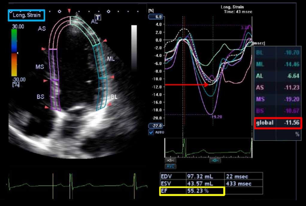
Other investigators have shown that myocardial deformation (strain imaging) with speckle tracking echocardiography (STE) holds considerable promise as a tool for predicting which patients will develop cardiotoxicity during or after cancer therapy. Reductions in echocardiographic measures of myocardial deformation parameters (strain and strain rate) are a sign of subclinical myocardial changes from cancer therapy and occur prior to any change in LVEF as assessed by conventional 2D echocardiography.

Importantly, early reduction in myocardial deformation appears to forecast the development of subsequent cardiotoxicity, with STE measured global longitudinal strain (GLS) being the most consistent parameter. The thresholds of change in GLS to predict cardiotoxicity have ranged from 10% to 15% using STE. An early fall in GLS by STE between 10% and 15% predicts subsequent cardiotoxicity (including both asymptomatic and symptomatic LV dysfunction).

Hence, there has been a growing interest in markers of early myocardial changes (i.e., changes with normal LVEF) that may predict the development of subsequent LVEF reduction or the progression to HF, so that preventive strategies with established cardioprotective medications such as beta-blockers, angiotensin-converting enzyme inhibitors, or dexrazoxane could be implemented.

When treatment with enalapril and carvedilol was initiated within the first couple of months following the end of chemotherapy, 64% of patients experienced complete recovery of their LVEF. When the heart failure medications were commenced 3-4 months after completing chemotherapy, the LVEF recovery rate dropped to 28%. No complete recovery of LVEF occurred in patients who began enalapril plus carvedilol after 6 months. Predictors of increased risk are helpful in identifying cancer therapy-related cardiotoxicity early in its course when aggressive treatment with standard heart failure medications such as beta-blockers and ACE inhibitors (ACEI) is most likely to be beneficial. Complete LVEF recovery and associated cardiac events reduction may be achieved when cardiac dysfunction is detected early and treatment with ACEI, possibly in combination with beta-blockers, is promptly initiated.

Source:

. Thavendiranathan P, et al. Use of Myocardial Strain Imaging by Echocardiography for the Early Detection of Cardiotoxicity in Patients During and After Cancer Chemotherapy: A Systematic Review. J Am Coll Cardiol. 2014;63(25_PA):2751-2768. doi:10.1016/j.jacc.2014.01.073

. Cardinale D, et al. Anthracycline-Induced Cardiomyopathy: Clinical Relevance and Response to Pharmacologic Therapy. J Am Coll Cardiol. 2010;55(3):213-220. doi:10.1016/j.jacc.2009.03.095


Myocardial Strain Imaging by Echocardiography for the Early Detection of Chemotherapy-induced Cardiotoxicity

The mortality rate among patients with cancer has decreased over the past 20 to 30 years. However, cardiac toxicity (cardiotoxicity) from cancer therapy has become a leading cause of morbidity and mortality in survivors. In patients who develop heart failure (HF) from cancer therapy, the mortality rate is as high as 60% by 2 years. Therefore, contemporary management of patients with cancer should include careful consideration of potential cardiotoxicity during therapy, with a focus on early detection and intervention.

Several definitions of cardiotoxicity have been proposed. The most commonly used definition is a ≥ 5% reduction in symptomatic patients (or ≥10% reduction in asymptomatic patients) in the left ventricular ejection fraction (LVEF) from baseline to an LVEF < 55%. Early detection of cardiotoxicity has predominantly relied upon serial cardiac imaging to identify a reduction in left ventricular (LV) function without signs or symptoms of heart failure (stage B HF).

The use of LVEF has important limitations. First, the measurement of LVEF is subject to technique-related variability, which can be higher than the thresholds used to define cardiotoxicity. Second, the reduction in LVEF is often a late phenomenon, with failure to recover systolic function in up to 58% of patients despite intervention. Hence, there has been a growing interest in markers of early myocardial changes (i.e., changes with normal LVEF) that may predict the development of subsequent LVEF reduction or the progression to HF, so that preventive strategies with established cardioprotective medications such as beta-blockers, angiotensin-converting enzyme inhibitors, or dexrazoxane could be implemented.

All studies of early myocardial changes with chemotherapy demonstrate that alterations of myocardial deformation precede significant change in left ventricular ejection fraction (LVEF). Using tissue Doppler-based strain imaging, peak systolic longitudinal strain rate has most consistently detected early myocardial changes during therapy, whereas with speckle tracking echocardiography (STE), peak systolic global longitudinal strain (GLS) appears to be the best measure. A 10% to 15% early reduction in GLS by STE during therapy appears to be the most useful parameter for the prediction of cardiotoxicity, defined as a drop in LVEF or heart failure.

A systemic review published in July 2014 in the American Journal of Cardiology (JACC), by P. Thavendiranathan et al.,  confirms the value of echocardiographic myocardial deformation (strain imaging) parameters for the early detection of myocardial changes and prediction of cardiotoxicity in patients receiving cancer therapy.

These results can be summarized into the following key points:

  1. Cardiotoxicity from cancer therapy is a leading cause of morbidity and mortality in cancer survivors, and is most typically defined as a decrease in left ventricular ejection fraction (LVEF) by ≥ 5% or ≥ 10% for symptomatic and asymptomatic patients, respectively. As early identification may alter management and attenuate cardiotoxicity, there is a need for early markers of cardiotoxicity before a significant change in EF occurs.
  2. Echocardiographic assessment of LVEF or diastolic function does not appear to be able to identify cardiotoxicity at an early subclinical phase.
  3. Existing studies consistently identify that changes in myocardial deformation happen earlier than changes in LVEF. Using two-dimensional strain imaging, global longitudinal strain is more reproducible than other measures of strain such as global radial strain or global circumferential strain. Using tissue Doppler-based imaging, longitudinal strain rate appears to be a consistent marker of changes in myocardial deformation, while other measures appear to be less reliable.
  4. On a review of the literature examining the prognostic value of strain imaging in chemotherapy patients, an early decrease of 10-11% (95% confidence interval, 8-15%) in global longitudinal strain predicts cardiotoxicity. Other individual markers have not been predictive, although combined use of global longitudinal strain and LV twist may be a better predictor than the former variable alone.
  5. Late cardiotoxicity can be observed several years after chemotherapy is completed. While there are multiple studies examining myocardial deformation during a longer-term follow-up after treatment, the relationship of abnormal findings and prognosis remains uncertain.
  6. Radiotherapy may also be associated with early changes of the myocardium, and some literature has observed a change in myocardial deformation immediately following treatment, although separating the effects from radiotherapy and chemotherapy is difficult, as they are often used simultaneously.
  7. Current recommendations for cardiac evaluation of patients prior to cancer treatment are variable and not specific. While multiple modalities can be used, echocardiography has advantages given its versatility, low cost, and excellent safety profile.
  8. While normal ranges for global longitudinal strain from a recent meta-analysis suggest a normal cutoff of -19% to -22%, there is significant between-patient variability, suggesting that within-patient changes in strain may be more reliable than population-based thresholds.
  9. While strain imaging is a promising method to identify early cardiotoxicity, further multicenter study is needed that includes cancers other than breast cancer, and compares management and outcomes between patients initiated on cardioprotective therapy on the basis of strain imaging versus traditional measures such as LVEF.

Source:

Thavendiranathan P, Poulin F, Lim KD, Plan JC, Woo A, Marwick TH. Use of Myocardial Strain Imaging by Echocardiography for the Early Detection of Cardiotoxicity in Patients During and After Cancer Chemotherapy – A Systematic Review J Am Coll Cardiol. 2014;63(25_PA):2751-2768. doi:10.1016/j.jacc.2014.01.073

LaBounty TM (Cardiosource).

2014 AHA/ACC Guideline for the Management of Patients with Non-ST-Elevation Acute Coronary Syndromes (NSTE-ACS): 10 Points to Remember

The new guideline emphasizes the pathophysiologic continuum of unstable angina and NSTEMI and their frequently indistinguishable clinical presentations. In terms of treatment, another notable element of the new guideline, is the recognition that while an early invasive strategy for patients with NSTE-ACS with significant coronary artery disease is generally accepted, low-risk patients can substantially benefit from guideline-directed medical therapy. Guideline-directed medical therapy has not always been optimally utilized and advances in noninvasive testing have the potential to identify patients with NSTE-ACS at low-intermediate risk to distinguish candidates for invasive versus medical therapy.

In addition, the guideline recognizes important, developing clinical areas requiring further research such as the utility of combined, potent antithrombotic and anticoagulant therapy in certain patient groups, appropriate application of new, high-sensitivity troponins, and the proper selection of high-risk elderly patients and women for interventional therapy. The guideline also contains expanded recommendations regarding discharge, such as education about symptoms, risk modification, routine medication with dual antiplatelet therapy, cholesterol management, referral to cardiac rehabilitation, and other guideline-directed medical therapy.

The following are 10 points to remember about this guideline:

1. Patients with suspected ACS should be risk stratified based on the likelihood of ACS and adverse outcome(s) to decide on the need for hospitalization and assist in the selection of treatment options.

2. In patients with chest pain or other symptoms suggestive of ACS, a 12-lead electrocardiogram (ECG) should be performed and evaluated for ischemic changes within 10 minutes of the patient’s arrival at an emergency facility.

3. Cardiac-specific troponin (troponin I or T with a contemporary assay) levels should be measured at presentation and 3-6 hours after symptom onset in all patients who present with symptoms consistent with ACS to identify a rising and/or falling pattern.

4. Oral beta-blocker therapy should be initiated within the first 24 hours in patients who do not have any of the following: 1) signs of heart failure, 2) evidence of low-output state, 3) increased risk for cardiogenic shock, or 4) other contraindications to beta blockade (e.g., PR interval > 0.24 second, second- or third-degree heart block without a cardiac pacemaker, active asthma, or reactive airway disease).

5. High-intensity statin therapy should be initiated or continued in all patients with NSTE-ACS and no contraindications to its use.

6. A P2Y12 inhibitor (either clopidogrel or ticagrelor) in addition to aspirin should be administered for up to 12 months to all patients with NSTE-ACS without contraindications who are treated with either an early invasive or ischemia-guided strategy. P2Y12 inhibitor therapy (clopidogrel, prasugrel, or ticagrelor) continued for at least 12 months is indicated in post–percutaneous coronary intervention (PCI) patients treated with coronary stents. It is reasonable to use ticagrelor in preference to clopidogrel for P2Y12 treatment in patients with NSTE-ACS who undergo an early invasive or ischemia-guided strategy. It is also reasonable to choose prasugrel (initiated during PCI) over clopidogrel for P2Y12 treatment in patients with NSTE-ACS who undergo PCI who are not at high risk of bleeding complications.

7. In patients with NSTE-ACS, anticoagulation, in addition to antiplatelet therapy, is recommended for all patients irrespective of initial treatment strategy. In patients with NSTE-ACS, anticoagulant therapy should be discontinued after PCI unless there is a compelling reason to continue such therapy.

8. An urgent/immediate invasive strategy (diagnostic angiography with intent to perform revascularization if appropriate based on coronary anatomy) is indicated in patients with NSTE-ACS who have refractory angina or hemodynamic or electrical instability (without serious comorbidities or contraindications to such procedures). An early invasive strategy (diagnostic angiography with intent to perform revascularization if appropriate based on coronary anatomy) is indicated in initially stabilized patients with NSTE-ACS (without serious comorbidities or contraindications to such procedures) who have an elevated risk for clinical events. An early invasive strategy (i.e., diagnostic angiography with intent to perform revascularization) is not recommended in patients with extensive comorbidities (e.g., hepatic, renal, pulmonary failure, cancer), in whom the risks of revascularization and comorbid conditions are likely to outweigh the benefits of revascularization and those with acute chest pain and a low likelihood of ACS who are troponin-negative, especially women.

9. All eligible patients with NSTE-ACS should be referred to a comprehensive cardiovascular rehabilitation program either before hospital discharge or during the first outpatient visit.

10. An evidence-based plan of care (e.g., guideline-directed medical treatment) that promotes medication adherence, timely follow-up with the healthcare team, appropriate dietary and physical activities, and compliance with interventions for secondary prevention should be provided to patients with NSTE-ACS. In addition to detailed instructions for daily exercise, patients should be given specific instruction on activities (e.g., lifting, climbing stairs, yard work, and household activities) that are permissible and those to avoid. Specific mention should be made of resumption of driving, return to work, and sexual activity.

 Source:

CardioSource

What Is a Normal ECG in Athletes?

Up to 60% of athletes show ECG changes, and some of these changes are often striking to look at.  The Seattle ECG criteria is often used to separate the normal from the abnormal ECG in an athlete. This is important, as many normal athletes undergo cardiac angiography and even pacemaker surgery because of misinterpretation of their ECGs.

  • Bradycardia: A low heart rate in an athlete should be self-evident—but it is not in the real world. Common normal athletic ECG patterns include: the inverted P-wave and junctional rhythm, Mobitz type I Wenckebach block, and sinus arrhythmia.
  • Incomplete right bundle branch block (IRBBB): While only 10% of the general population has IRBBB, up to 40% of athletes have it as a normal pattern. This might be due to remodeling of the athletic right ventricle.
  • Left ventricular hypertrophy (LVH): It is normal for athletes to have voltage criteria for LVH. Of course, it is abnormal when voltage criteria comes with other LVH related patterns, such as left atrial abnormality, ST-segment strain patterns, left axis deviation, etc.
  • Early repolarization: It is common and normal for athletes to have early repolarization, including terminal QRS notching. The pattern is especially common in black athletes.
  • T-wave changes: T-wave inversions can be normal, or they can be abnormal. Up to one in three black athletes have terminal T-wave inversions in leads V1–V4. T-wave inversion patterns which can be mistaken for anterior-wall ischemia. Paying attention to the location of the inversions, however, is important. Namely, it is normal to have T-wave inversions in leads V1–V4 but not in V5 or V6 and not with accompanying QRS abnormalities or frequent premature ventricular contractions (PVCs).
  • Notable abnormal findings in athletes: Some of the abnormal ECG findings may include any complete left bundle branch block, intraventricular conduction delay (IVCD) greater than 140 ms, left atrial abnormality, right ventricular hypertrophy, Wolff-Parkinson-White (WPW) syndrome, QT prolongation (greater than 470 ms in males and 480 ms in females), high-degree AV block, atrial fibrillation/flutter, and high-density PVCs. Isolated (low-density) PVCs are considered benign.


Colchicine for Recurrent Pericarditis

In a new trial (Lancet March 30, 2014), colchicine, was found to be safe and beneficial adjunct to nonsteroidal anti-inflammatory drugs, even in patients with multiple recurrences.  Previous trials have demonstrated the efficacy and safety of colchicine as an adjunct to conventional treatment in patients with acute pericarditis or first recurrences.

To assess whether colchicine’s effectiveness extends to multiple recurrences, investigators in Italy conducted the multicenter, double-blind, CORP-2 trial. They randomly assigned 240 patients with ≥ 2 recurrences of pericarditis to receive placebo or 0.5 mg colchicine (twice daily in patients heavier than 70 kg; once daily in those ≤70 kg) for 6 months. All participants also received nonsteroidal anti-inflammatory drugs (NSAIDs) or corticosteroids.                                                 During a mean follow-up of 20 months, the rate of pericarditis recurrence was about half as high in the colchicine group as in the placebo group (21.6% vs. 42.5%; P<0.001), and the rate of remission at 1 week of treatment was significantly higher in the colchicine group (83.3% vs. 59.2%). Rates of adverse events and drug discontinuation were low and similar in both groups.

These findings suppot the evidence that colchicine is a safe and effective adjunct to NSAIDs for the treatment of pericarditis.

Source:

Imazio M et al. for the ICAP Investigators. A randomized trial of colchicine for acute pericarditis. N Engl J Med 2013 Sep 1; [e-pub].                                                                                                                                                                     

Imazio M et al. Efficacy and safety of colchicine for treatment of multiple recurrences of pericarditis (CORP-2): A multicentre, double-blind, placebo-controlled, randomised trial. Lancet 2014 Mar 30; [e-pub].


β blockers for Heart Failure: Which One Should You Use?

β blockers are a cornerstone of the medical management of heart failure. The long term use of certain β blockers in patients with heart failure reduces hospital admissions and improves symptoms, quality of life, and survival. However, it is still unclear whether this is a class effect or whether one β blocker is superior to another.    

Three landmark placebo-controlled trials of nearly 9000 patients with heart failure demonstrated the efficacy of carvedilol, long acting metoprolol succinate, and bisoprolol in reducing mortality and hospital admission in patients with heart failure. However, large trials of nebivolol and bucindolol found no reductions in all-cause mortality compared with placebo, despite benefits in cardiovascular morbidity or mortality (1).

A systematic review and meta-analysis of 8 randomized, controlled, direct-comparison trials involving 4,563 patients with systolic heart failure receiving atenolol, bisoprolol, metoprolol, nebivolol, or carvedilol.  In this analysis, carvedilol, as compared against atenolol, bisoprolol, metoprolol and nebivolol in randomized direct comparison trials, significantly reduced all-cause mortality in systolic HF patients (2).

A more recent meta-analysis analyzed the results of 21 randomized trials (focusing on atenolol, bisoprolol, bucindolol, carvedilol, metoprolol, and nebivolol) comparing β blockers with other β blockers or other treatments in patients with heart failure and reduced ejection fraction (3). The primary endpoint was all cause death at the longest available follow-up, assessed with odds ratios and Bayesian random effect 95% credible intervals, with independent extraction by observers.   As expected, in the overall analysis, β blockers provided credible mortality benefits in comparison with placebo or standard treatment after a median of 12 months (odds ratio 0.69, 0.56 to 0.80). However, no obvious differences were found when comparing the different β blockers head to head for the risk of death, sudden cardiac death, death due to pump failure, or drug discontinuation. Accordingly, improvements in left ventricular ejection fraction were also similar irrespective of the individual study drug.

CONCLUSION: The benefits of β blockers in patients with heart failure with reduced ejection fraction seem to be mainly due to a class effect, as no statistical evidence from current trials supports the superiority of any single agent over the others. Therefore, β blockers reduce mortality but do not differ from each other.  

Effect of ACE Inhibitors and ARBs on Cardiovascular Outcomes in Diabetes

A recent meta-analysis, published in JAMA Intern Med on March 31, 2014, analyzed the effect of ACE inhibitors (ACEIs) and angiotensin II receptor blockers (ARBs) on all-cause mortality, cardiovascular (CV) deaths, and major CV events in patients with diabetes mellitus (DM). The authors of this study included randomized clinical trials reporting the effects of ACEI and ARB regimens for DM on all-cause mortality, CV deaths, and major CV events with an observation period of at least 12 months.

The major results of 35 trials involving more than 50,000 patients showed that ACEIs reduced all-cause mortality, CV mortality, and major CV events in patients with DM.

By contrast, ARBs did not reduce all-cause mortality, CV mortality, or major CV events. Neither ACEIs nor ARBs were associated with a decrease in the risk for stroke in patients with DM. This study suggests that ACEIs vs ARBs be used as first-line therapy in patients with diabetes.  Primary end points were all-cause mortality and death from CV causes. Secondary end points were the effects of ACEIs and ARBs on major CV events.

Twenty-three of 35 identified trials compared ACEIs with placebo or active drugs (32,827 patients) and 13 compared ARBs with no therapy (controls) (23 867 patients). When compared with controls (placebo/active treatment), ACEIs significantly reduced the risk of all-cause mortality by 13%, CV deaths by 17%, and major CV events by 14%, including myocardial infarction by 21% and heart failure by 19%.

Treatment with ARBs did not significantly affect all-cause mortality, CV death rate, and major CV events with the exception of heart failure.

Both ACEIs and ARBs were not associated with a decrease in the risk for stroke in patients with DM. Meta-regression analysis showed that the ACEI treatment effect on all-cause mortality and CV death did not vary significantly with the starting baseline blood pressure and proteinuria of the trial participants and the type of ACEI and DM.

The authors concluded that angiotensin-converting enzyme inhibitors reduced all-cause mortality, CV mortality, and major CV events in patients with DM, whereas ARBs had no benefits on these outcomes. Thus, ACEIs should be considered as first-line therapy to limit excess mortality and morbidity in this population.

Source:

Cheng J, Zhang W, Zhang X, et al. Effect of Angiotensin-Converting Enzyme Inhibitors and Angiotensin II Receptor Blockers on All-Cause Mortality, Cardiovascular Deaths, and Cardiovascular Events in Patients With Diabetes Mellitus: A Meta-Analysis JAMA Intern Med 2014 Mar 31;[EPub Ahead of Print].

Antibiotics Linked to Increased Risks for Arrhythmia and Mortality

Azithromycin use has been associated with increased risk of death among patients at high baseline risk, but not for younger and middle-aged adults. The Food and Drug Administration issued a public warning on azithromycin, including a statement that the risks were similar for levofloxacin. Researchers conducted a retrospective cohort study among US veterans to test the hypothesis that taking azithromycin or levofloxacin would increase the risk of cardiovascular death and cardiac arrhythmia compared with persons taking amoxicillin.

Azithromycin and levofloxacin carry higher arrhythmia and mortality risks than amoxicillin, according to an observational study in the Annals of Family Medicine. The FDA issued a cardiac warning on azithromycin in March 2013.  Researchers studied nearly 1.8 million U.S. veterans (mean age, 57) who received outpatient prescriptions for one of the three antibiotics from 1999 to 2012.    On weighted analysis, the numbers of deaths by day 5 of treatment per million antibiotics dispensed were 154 for amoxicillin, 228 for azithromycin, and 384 for levofloxacin.  Patients receiving azithromycin had roughly a 50% increased risk for death and an 80% increased risk for serious arrhythmia, compared with those on amoxicillin. Risk increases were even greater for levofloxacin.

The researchers conclude: “There are usually multiple antibiotic choices available for older patients, especially those with cardiac comorbidities; physicians may consider prescribing medications other than azithromycin and levofloxacin.”

New (2014) AHA/ACC Guideline for Management of Valvular Heart Disease

New, 2014 AHA/ACC, Guideline   for the Management of Patients With Valvular Heart Disease: has been   released.  This document is   a major revision of the 2006 and 2008 American College of Cardiology   (ACC)/American Heart Association (AHA) guidelines on the management of   patients with valvular heart disease. There are substantial changes in   multiple areas, and practitioners who are involved in the care of patients   with heart valve disease should read at least the executive summary, if not   the full guideline document.

The following are 10 areas with important changes to remember:

1. Stages of heart valve disease. The revised guidelines include a new classification of heart valve diseases with four progressive stages: A, at risk; B, progressive; C, asymptomatic severe; and D, symptomatic severe. For each valve lesion, stages are based on valve anatomy, valve hemodynamics, hemodynamic consequences, and symptoms.

2. The heart valve team. The guidelines recommend (Class I) that the management of patients with severe heart valve disease is best achieved by a heart valve team, composed minimally of a cardiologist and a cardiac surgeon; but potentially including cardiologists, structural valve interventionalists, cardiovascular imaging specialists, cardiovascular surgeons, anesthesiologists, and nurses, all of whom have expertise in the management and outcome of patients with severe heart valve disease.

3. Heart valve centers of excellence. The guidelines recommend (Class IIa) that consultation with or referral to a heart valve center of excellence is reasonable for asymptomatic patients with severe valve disease, patients with disease that could be best treated with valve repair rather than replacement, and patients with multiple comorbidities in whom valve intervention is considered. Specific criteria for a heart valve center of excellence include experienced providers from multiple disciplines, an ability to offer all available options for diagnosis and management, participation in regional or national outcome registries, demonstrated adherence to guidelines, ongoing quality improvement processes, and public reporting of intervention mortality and success rates.

4. Evaluation of surgical and interventional risk. The guidelines provide specific recommendations for the assessment of surgical and interventional risks that include the Society of Thoracic Surgeons (STS) predicted risk of mortality (PROM) calculator, patient frailty (e.g., Katz Activities of Daily Living and independence), the number of compromised major organ systems, and procedure-specific impediments.

5. Exercise testing. The guidelines place increased emphasis (Class IIa) on the use of exercise testing in the evaluation of asymptomatic patients with severe heart valve disease (notably including asymptomatic severe aortic stenosis [AS] and asymptomatic severe primary mitral regurgitation [MR]); aimed at confirmation of the absence of symptoms, assessment of the hemodynamic response to exercise, and assessment of prognosis.

6. Aortic stenosis. Changes were made in the characterization of and indications for intervention for AS:

  • Two important changes were made in the characterization of AS:

i)    ‘very severe’ AS is defined as aortic Vmax ≥5 m/s or mean gradient ≥60 mm Hg; and

ii)   symptomatic severe AS is subdivided into high gradient (Vmax ≥4 m/s or mean gradient ≥40 mm Hg), low-flow low-gradient (LFLG) with reduced left ventricular ejection fraction (LVEF) (severe leaflet calcification with severely reduced motion, effective orifice area [EOA] ≤1.0 cm2, and Vmax <4 m/s or gradient <40 mm Hg with LVEF <50%, and EOA remaining ≤1.0 cm2, but Vmax ≥4 m/s at any flow rate during dobutamine echocardiography), and LFLG with normal LVEF (or paradoxical LFLG severe AS; severe leaflet calcification with severely reduced motion, EOA ≤1.0 cm2 and Vmax <4 m/s or gradient <40 mm Hg with LVEF ≥50%).

  • Indications for intervention are expanded from previous to include patients with very severe AS (above) and low surgical risk (Class IIa); asymptomatic severe AS and decreased exercise tolerance or exercise-related decrease in blood pressure (Class IIa); and symptomatic  patients with LFLG severe AS and normal LVEF if clinical, hemodynamic, and  anatomic data support valve obstruction as the likely cause of symptoms      (Class IIa).

7. Transcatheter aortic valve replacement. Surgical aortic valve replacement (AVR) remains the intervention of choice for patients with an indication for AVR and low or intermediate operative risk (Class I). Transcatheter AVR (TAVR) is recommended for patients with an indication for AVR, but a prohibitive surgical risk (Class I). TAVR is a reasonable alternative to surgical AVR in patients with an indication for AVR and high surgical risk (Class IIa). Notably, members of a heart valve team should collaborate in order to optimize patient care for patients in whom TAVR or high-risk surgical AVR are being considered (Class I).

8. Management of primary MR. A clear distinction is drawn between chronic primary (degenerative) MR, with pathology of one or more components of the valve (leaflets, annulus, chordae, papillary muscles); and chronic secondary (functional) MR. Intervention for severe chronic primary MR remains indicated (all Class I) for symptoms, LV dysfunction (LVEF ≤60% and/or systolic diameter ≥40 mm), and at the time of other cardiac surgical intervention. Changes in the recommendations for mitral valve repair (MVr) include the following:

  • MVr is recommended over mitral valve replacement (MVR) when pathology  is limited to the posterior leaflet (Class I);
  • MVr is recommended over MVR when pathology involves the anterior or both leaflets and a successful and durable repair can be accomplished (Class I);
  • ‘Prophylactic’ MVr (repair in an asymptomatic patient with preserved LV function) is reasonable when performed at a heart valve center of excellence and the likelihood of successful and durable repair without      residual MR is >95% and operative mortality risk is <1% (Class IIa);
  • MVr is reasonable in an asymptomatic patient with severe nonrheumatic MR and preserved LV function in the setting of new-onset atrial fibrillation or resting pulmonary artery systolic pressure >50 mm Hg (Class IIa); and
  • MVr may be considered in the setting of rheumatic mitral valve disease when surgery is indicated and either successful repair is likely or when long-term anticoagulation management appears unreliable (Class IIb). There      is a Class III (harm) indication for MVR in the setting of isolated disease involving less than half of the posterior mitral leaflet unless repair was attempted and was unsuccessful.
  • Finally, transcatheter MVr may be considered in severely symptomatic patients who have favorable anatomy and a reasonable life expectancy and a prohibitive surgical risk due to comorbidities, and remain severely      symptomatic despite optimal medical therapy (Class IIb).

9. Management of secondary MR. Chronic secondary (functional) MR occurs as a result of abnormalities of the LV, and has more differences than similarities with chronic primary MR (above). The treatment of chronic secondary MR involves treatment of the underlying cardiomyopathy (Class I) and cardiac resynchronization therapy if indicated (Class I). Intervention for chronic secondary MR is reasonable at the time of coronary artery bypass grafting or other cardiac surgery if MR is severe (Class IIa) and not unreasonable if MR is moderate (Class IIb). Surgical intervention performed primarily for chronic secondary MR remains limited to severely (New York Heart Association class III-IV) symptomatic patients with persistent symptoms despite optimal medical therapy for heart failure (Class IIb); new in these guidelines is acceptance of equivalence among these patients between MVR and MVr.

10. Low molecular weight heparin (LMWH) ‘bridging’ of patients with a mechanical heart valve prosthesis. Low LMWH ‘bridging’ is now considered to be appropriate (Class I) among patients with a mechanical valve at high risk of thrombosis when warfarin needs to be interrupted in the context of an invasive or surgical procedure (previously Class IIb).


Source: D.S. Bach, MD (Cardiosource)