Echocardiogram
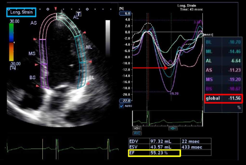
The operator puts some clear gel on your chest and then places an ultrasound probe on it. The probe sends ultrasound beams into your body and their reflections are detected and used to generate images of the heart. You can see different parts of your heart on a screen as the probe is moved around on your chest.
(Also called an ‘echo’.)
This test uses ultrasound waves to look at the structure of the heart. It is useful for people whose ECG shows changes that could be caused either by a channelopathy or by uninherited heart disease that has damaged the heart – for example a previous heart attack that you may not have even been aware of. An echocardiogram can also detect inheritable conditions such as cardiomyopathy and mitral valve prolapse.
**Source: sads.org.uk


