About the Heart’s Electrical System
About the Heart’s Electrical System
When most people think of the heart, they think of the arteries of the heart, or of the heart muscle and valves. But the heart also has a complex electrical system that coordinates the rhythm. Abnormalities in the heart’s electrical system can lead to arrhythmias – causing the heart to beat too fast or too slowly.
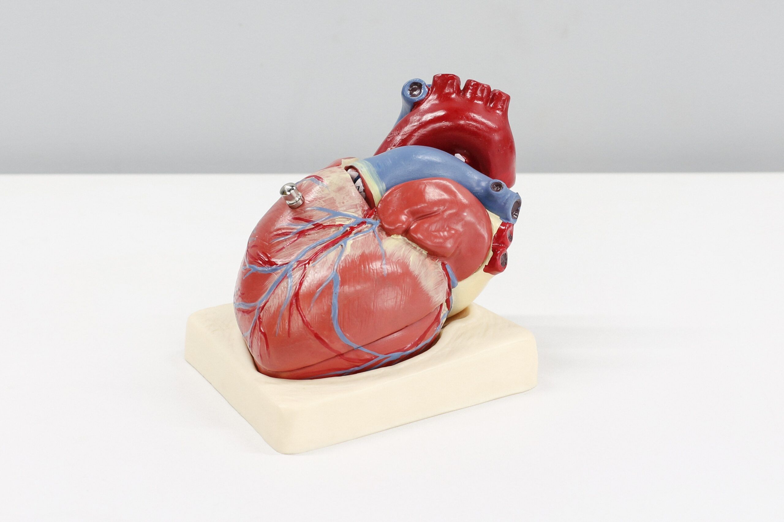
The heart is divided by muscle and fibrous tissue into a right and left side. Each side has an upper chamber or atrium that collects blood returning to the heart and a muscular lower chamber or ventricle that pumps that blood away from the heart. The right atrium (RA) receives blood from you body and pumps it into the right ventricle (RV). The right ventricle then pumps it to your lungs. From the lungs, blood returns to your left atrium (LA) and then pumped into the left ventricle (LV). From the left ventricle it is pumped out to your body.
 To make sure that the different parts of the heart work together and pump the blood with the right timing and sequence, the heart uses its built-in “electrical system”. Think of it as a set of nerves or wires that run throughout the heart connecting all the parts. In the right atrium you will find the sinoatrial (SA) node – the “pacemaker” of the heart. It sends electrical impulses just like a spark plug in a car. The SA node thus sets your heart rate. This impulse spreads throughout the atria like ripples in a pond, and then travels down to what we call the atrioventricular (AV) node. The AV node is similar to a wire or cable connecting the atria and the ventricles and is responsible for sending electrical impulses from the atria to the ventricles. It splits into 2 branches called the right and left bundle branch which allows the even spread of the electrical signal to both ventricles simultaneously.
Specific Rhythm Problems Wolff-Parkinson-White (WPW) Syndrome
In the normal heart, the AV node is the only electrical connection between the atria and ventricles. With WPW, the heart has an extra nerve, pathway or wire which we call an “accessory pathway” that electrically connects the atrium to the ventricle. It is present from birth but may not be detected or cause any problems with tachycardia until later in life. Many WPW patients may not ever experience heart problems from this abnormal nerve and may not even know they have it until it is detected by a doctor. This pathway is in the wall of the heart and can be located anywhere on the right, left, front or back walls. Some symptomatic patients have what is called manifest preexcitation (this is true WPW) while others have what is called a concealed pathway (meaning that it cannot be easily seen on a resting ECG). Both manifest preexcitation and concealed pathways can cause troublesome arrhythmias – usually supraventricular tachycardia (SVT).
 Some people have more than one accessory pathway. People with WPW may experience tachycardia attacks because the electrical impulse gets trapped in an electrical circuit that travels in a large circle between the normal AV node and the accessory pathway. This can cause the heart to race up to 150 – 300 beats per minute. The tachycardia attacks start suddenly without warning and there is often no obvious cause. The feeling of the heart pounding in the chest or neck can be associated with lightheadedness, chest pain and sometimes a blackout. Rarely, WPW can cause the heart to race rapidly and dangerously out of control. Catheter ablation of WPW syndrome involves destroying the accessory pathway. The EP study determines the number of extra pathways and the location of each. The ablation catheter is then inserted and used to carefully map the heart to precisely locate each pathway. Each time that the catheter locates an extra pathway and is placed against it, ablation energy is delivered. AV Node Reentrant Tachycardia (AVNRT)
Another common problem that can cause supraventricular tachycardia (SVT) is AV node reentry. This rhythm problem is due to an abnormality of the AV node itself which results in a “short circuit” in the area around the AV node. An electrical signal can get trapped into a small loop in this area, causing the heart to race. The tachycardia may feel very similar to that experienced by those with WPW syndrome simply because a racing heart still feels like a racing heart regardless of the cause.
 Catheter ablation is directed at destroying the tissue near the AV node causing the heart racing without causing serious damage to the AV node. To minimize this risk, your doctor will start by burning a safe distance from the AV node and gradually burn tissue progressively closer until he/she sees signs that enough burning has been done or if signs of elevated risk appear. The technique is similar to whittling away at a piece of wood where you shave away part of the wood without weakening it and causing it to break. Unfortunately, the abnormal tissue can be very, very close to the AV node and damage to the AV node occurs in a small percentage of cases despite all measures taken to avoid it. If this should happen, the implant of a pacemaker may be necessary.
Atrial Flutter
In atrial flutter, the electrical signal gets trapped in a loop running around the right atrium, causing it to beat at 300 beats per minute while the ventricles beat usually at 150 beats per minute. The abnormal tissue causing this rhythm problem is located near the bottom of the right atrium. This area is easy to reach with the ablation catheter but it may be thick and uneven, making it somewhat difficult to burn all the tissue necessary to eliminate the flutter.
 It is also important to point out that people with atrial flutter can have more than one kind of flutter – so ablation sometimes has to be directed to different places in the heart. Many patients with atrial flutter will also have attacks of atrial fibrillation as well, which is a related but totally different arrhythmia that requires a different slate of treatment strategies. Eliminating atrial flutter alone in patients with both atrial flutter and atrial fibrillation may not reduce or eliminate atrial fibrillation attacks. Patients who have continued problems with attacks of atrial fibrillation after a successful flutter ablation may be candidates for other types of ablation procedures.
Atrial Fibrillation
Atrial fibrillation (often called “A Fib”) is most commonly seen in people who have other heart disease (such as heart valve problems, heart attacks, long-standing high blood pressure) or thyroid disease but can be seen in otherwise healthy people without any medical problems. The atria become scarred and irritable and are not able to pass the electrical impulse smoothly like a ripple traveling across a calm water pond. Instead, the electrical impulse breaks up into many smaller ripples that travel around the atria in a very fast, irregular and disorganized manner much like a stormy ocean surface. This makes the atria beat at between 300-600 beats/minute. A proportion of these impulses travel down the AV node and cause the ventricles to beat quite fast (120-190 beats/min) and very irregularly. The atria beat so fast that blood does not get pumped normally and blood clots may form in the atria. Therefore, blood thinners are often prescribed. Atrial fibrillation is often very difficult to control with drugs.
 A number of different drugs are used to treat atrial fibrillation. Some drugs are prescribed to try and prevent atrial fibrillation attacks from recurring. Other drugs (beta blockers, calcium blockers, digoxin) are used to slow the heart when atrial fibrillation occurs and makes the attacks more tolerable or less uncomfortable but do not prevent the attacks from recurring. When a person cannot tolerate medications or when drugs are not effective at preventing attacks or reducing symptoms from attacks, catheter ablation may be necessary.
There are two types of ablation that can be performed for atrial fibrillation. Each has a different approach and a different goal.
1) AV node-His bundle ablation: This type of ablation was the first type ever to be performed and was introduced in 1983. This treatment does not cure someone of AF attacks. Rather, it eliminates symptoms by destroying the AV node-His bundle so that the atrial fibrillation signals cannot cause the ventricles to beat rapidly and irregularly. After the ablation, AF is still present BUT people no longer have any symptoms from the fibrillation. AV node ablation is 99% successful on the first attempt and the risks are very low (<1%). Remember however, that before your AV node ablation, a permanent pacemaker will be required. This may be done on the same day or some weeks in advance of your AV node ablation. It is best to think of AV node ablation as an ablation that necessarily requires a pacemaker implant as a part of the treatment strategy.
2) AF ablation (also known as Pulmonary Vein Ablation (PVA): In recent years, new research has found that many patients have atrial fibrillation that is triggered by one or more spots in the atrial chambers. Heart cells in these areas send out rapid electrical pulses and start the atrial fibrillation just like a malfunctioning ignition on a gas barbeque or oven. The most common site for these abnormal rapidly firing cells is in the pulmonary veins that connect to the left atrium. Pulmonary veins are veins that carry blood back from the lungs to the heart. Every person usually has four pulmonary veins but the most common veins causing atrial fibrillation are the two (left and right) upper veins. In focal atrial fibrillation, these spots are either destroyed by burning them or burning completely around the spots so they are trapped and the impulses coming from these cells are prevented from getting out to the rest of the heart and causing atrial fibrillation. The procedure can be very long (6 hours or more) and the success rate tends to be lower than that for catheter ablation for WPW, AVNRT or atrial flutter. It is also important to know that many (up to 50%) of these patients may require a second procedure – even when the first procedure has gone well.
This procedure is relatively new and is still being improved. Our knowledge about this procedure is advancing rapidly. At the present time, patients who have the highest success rates and the lowest complications are those who: 1) have otherwise normal hearts free of any scarring or damage from other heart disease, 2) have atrial fibrillation attacks that stop on their own (have periods where the heart beat returns to normal in between attacks and 3) have few other medical problems.
Ventricular Tachycardia
Ventricular tachycardia (often called VT) is a form of heart racing that starts inside the right or left ventricle. It is most commonly due to damage or scarring of the lower heart chambers, usually as a result of a previous heart attack. However, any disease that damages the heart can cause ventricular tachycardia to occur later in life. The time period between when the heart damage occurs and when ventricular tachycardia first develops can be days, months or up to many years. In someone who has heart disease, ventricular tachycardia is considered a potentially dangerous arrhythmia that requires careful treatment. Rarely, ventricular tachycardia can occur in healthy, young individuals without any history of heart disease (called idiopathic or primary VT). In these people, VT is NOT a dangerous problem but is nonetheless bothersome. With ventricular tachycardia, the heart can race at 130-250 beats per minute. Often, lightheadedness and blackouts can accompany the feeling of palpitations. Chest discomfort and shortness of breath may also be experienced.
 In patients whose VT is caused by previous damage or scarring of their hearts, catheter ablation is not often a good first choice for treatment. The implantation of an implantable cardioverter defibrillator (ICD for short) is preferred, along with medical therapy with heart-protective drugs like beta blockers, ACE inhibitors, and statins. Ablation therapy may be used in conjunction with the others to help reduce the frequency of attacks.
In VT patients who otherwise have normal hearts, catheter ablation is an alternative to drug therapy and can be curative. However, VT ablation can be more difficult because it may be difficult to turn on the VT at the time of the EP study. If the VT cannot be triggered, it is impossible to map and ablation cannot be done. This can happen in 25-40% of patients. The risks of VT ablation are usually less than 1-3%.
** Source: Canadian Heart Rhythm Society


